

Leigh A. Nelson, PharmD, BCPP

https://pharmacy.umkc.edu/directory/leigh-anne-nelson/
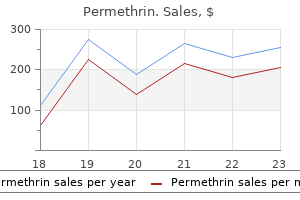
Permethrin dosages: 30 gm
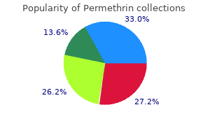
Permethrin packs: 3 creams, 4 creams, 5 creams, 6 creams, 7 creams, 8 creams, 9 creams, 10 creams

Recurrences are generally detected many years after primary remedy, and recurrent illness can be slowly progressive. Recurrent tumor could be borderline serous tumor, low-grade serous carcinoma or, hardly ever, high-grade serous carcinoma. Borderline serous tumors are large, usually multilocular cystic neoplasms which are bilateral in 35% to 40% of circumstances. Papillary growth is focal in some tumors, confluent in others, and present on the external surface of the ovary in 40% to 50% of instances. Areas of solid growth are unusual except in adenofibromatous borderline tumors, and zones of hemorrhage or necrosis are seldom seen. At low magnification, papillae with a hierarchical branching pattern grow from the cyst lining into the lumina. Complex papillary and glandular patterns and secondary cyst formation are typical. The papillae have fibrovascular cores that are conspicuous even in smaller branches and are surfaced by proliferating columnar cells stratified into a number of layers. Focally, the cells kind tufts from which clusters of cells and single cells are detached into the cyst lumen. Variable, but often low-grade, nuclear atypia and scattered mitotic figures are present. Cells with ample eosinophilic cytoplasm, the detached or metaplastic cells, are scattered singly or in small clusters among the many columnar tumor cells; they tend to be most conspicuous on the suggestions of the papillae. They resemble desmoplastic peritoneal implants (see later discussion) and are termed autoimplants. Some tumors have enough fibrous stroma in their partitions to be categorised as a borderline serous adenofibroma or cystadenofibroma. The microscopic characteristic that differentiates a serous borderline tumor from a serous carcinoma is the absence of diffuse stromal invasion within the former. In a borderline tumor, papillae and glands that seem to be within the stroma are an artifact resulting from tangential slicing of sophisticated infoldings of the cyst lining. Foci of restricted stromal invasion are recognized occasionally in a borderline serous tumor. Various arbitrary dimension limits have been proposed for microinvasion, ranging from three to 5 mm, but in practice foci of microinvasion are virtually always smaller than three mm.
By electron microscopy, the cytoplasm is seen to be filled with mitochondria, and other organelles are scant. Differential Diagnosis the principal consideration is the eosinophilic variant of chromophobe renal cell carcinoma. In most circumstances, strict adherence to the criteria listed in Table 12A-2 will allow this distinction. Cells with plentiful eosin ophilic cytoplasm are clustered in islands in an edematous stroma. Ultrastructurally, the cytoplasm of oncocytoma cells is filled with mitochondria, and other organ elles are scanty. Since that time, more than one hundred instances have been described individually or in aggregated research, and the name metanephric adenoma has turn out to be accepted. However, Beckwith now favors the name metanephric adenofibroma for these tumors to emphasize their close relationship with metanephric adenoma. Metanephric adenoma occurs at all ages, most commonly in the fifth and sixth a long time, and a 2: 1 female preponderance is seen. Four of the 50 sufferers reported by Davis and colleagues27 also had renal cell carcinoma. Patients with metanephric adenofibroma have ranged from thirteen months to 36 years (median 28 months). Other symptoms of metanephric adenofibroma have included hematuria26 and hypertension. Three of the five instances reported by Hennigar and Beckwith26 had separate small papillary epithelial tumors near the renal pelvis, which they thought-about low-grade accumulating duct carcinomas. Macroscopic Appearances Metanephric adenomas have ranged extensively in dimension with the biggest being one hundred fifty mm in diameter; most have been 30 to 60 mm in diameter. Small cysts are current in about 10% of tumors, and a unique instance was entirely cystic. Histologic Appearances Histologically, metanephric adenoma is typically a extremely cellular tumor composed of tightly packed small, uniform, spherical acini. Because the acini and their lumens are so small, at low magnification this sample may be mistaken for a stable sheet of cells. Hyalinized scar or focal osseous metaplasia of the stroma is present in 10% to 20% of tumors.
Diseases
The incidence of adenocarcinoma is quite variable amongst races and in several international locations, with the best reported incidence in North America, Australia, and Scandinavian countries. Prostate cancers are relatively rare in Asian populations, but recent information indicate that the incidence is rapidly rising. A geographic difference of incidence exists in Europe, with the next incidence in the international locations of northern and western Europe and lower in the nations of eastern and southern Europe. In addition to geographic, ethnic, and racial variations, genetic components are answerable for the completely different incidence as properly. Adenocarcinoma continues to be a fantastic scientific paradox; for example, in spite of the excessive incidence, extra males will die with adenocarcinoma somewhat than from the cancer itself. Autopsy research have shown that the incidence of adenocarcinoma increases with age. The majority are multifocal (60%-90%)80 and exhibit an acinar or combined acinar and ductal growth sample. Carcinomas could arise in any zone of the prostate, but the relative distribution is different in every zone; 68% of the carcinomas arise in the peripheral zone, 24% in the transition zone, and 8% in the central zone. The relatively low fatality compared with the high incidence of prostate cancer advised that adenocarcinomas are comparatively indolent and sufferers survive a lengthy time after the prognosis. Local unfold of adenocarcinoma occurs by way of extraprostatic extension or seminal vesicle invasion. Distant metastatic spread happens when carcinoma invades into lymphovascular areas. The most common websites of metastasis are regional pelvic lymph nodes, bone, and lung. Metastasis to the testis is rare, and unilateral involvement is extra common than bilateral involvement. First is the separation of well-differentiated adenocarcinoma from the vast variety of benign or atypical small-gland proliferations. Second is the edge for recognizing extraordinarily small foci of cancer in needle biopsies. Finally, on the excessive of the histologic spectrum, depending on the morphology, a very poorly differentiated adenocarcinoma of the prostate may be tough to distinguish from inflammatory infiltrates, metastatic carcinoma, and urothelial carcinoma involving the prostate. The principal criteria for analysis of welldifferentiated adenocarcinoma include a small-gland proliferation acknowledged as being discrete or focally infiltrative on low-power examination, the presence of a single cell lining with full absence of the basal cell layer, nucleomegaly, and presence of enormous nucleoli.
Entrapment of glomeruli and renal tubules is Congenital Mesoblastic Nephroma Although comprising fewer than 3% of main renal tumors in youngsters, congenital mesoblastic nephroma predominates within the first 3 months of life and is actually unknown after 24 months. The tumor was first acknowledged in 1966,317 and subsequent studies318 have shown this to be a morphologically distinct and prognostically favorable tumor. At the border between the tumor and the kidney, the spindle cells of the tumor infiltrate the interstitium between the renal tubules and glomeruli. In the cel lular variant, the tumor is more densely cellular, and the cells are round quite than spindle shaped. The cells have regular spherical to oval nuclei with chromatin clumping and vari able amounts of pale cytoplasm and are distributed in a matrix of dense bone. Another, and extra widespread, pattern was acknowledged later, consists of a more densely mobile proliferation of polygonal cells with easy-to-find mitotic figures, and infrequently has pushing borders. Some reports have instructed that the cellular sample is prone to recurrence, but, as noted above, age and completeness of resection appear to be the first threat factors for antagonistic outcome. Differential Diagnosis Mesoblastic nephroma usually is definitely diagnosed when the histopathology and patient age are considered. Wilms tumors with stromal predominance could also be confused with mesoblastic nephroma, particularly within the case of Wilms tumors treated before surgery. Although both occur in the identical age group, mesoblastic nephroma, even the mobile variant, and rhabdoid tumor are usually easily distinguished. Grossly, the tumors are sometimes stone-hard and project into the lumen of the renal pelvis. Microscopically, the majority of the tumors consists of sparsely mobile calcified osteoid containing nests of cells with small vesicular ovoid nuclei. These plump cells are bigger than osteocytes and are extra populous at the periphery of the lesion. Structures not ordinarily found in Wilms tumors, such as lymph node or gut-like buildings combining epithelium with clean muscle333 or hair follicle and sweat glands,334 are most useful. Delahunt B, Eble J N 1997 Papillary renal cell carcinoma: a clinicopathologic and immunohistochemical examine of a hundred and five tumors. J Urol 140: 721-724 Ossifying Renal Tumor of Infancy this uncommon tumor of unsure histogenesis has been reported in fewer than 20 patients, all youthful than 6 months. Although an irregular infiltrative border is current, the medical course has been benign to date.
Splenic Hemangioma and Hemangiomatosis Hemangioma is the most common major benign neoplasm of the spleen. A, Plump spindle cells with giant irregular-shaped, vesicular nuclei and vague cell borders are masked by a heavy lymphoplasmacytic infiltrate. These cells are shown on immunohistochemical studies to symbolize follicular dendritic cells. Whereas some spindle cells are relatively bland trying, others have overtly pleomorphic nuclei. The minimize surface of the spleen reveals multifocal lesions comprising aggregates of huge vascular areas. Peliosis of the spleen can be distinguished from hemangioma by the haphazard distribution of the irregular or round blood-filled areas with out formation of a discrete mass and the preferential involvement of the parafollicular areas. Histologically, anastomosing slim vascular channels are interspersed with dilated vascular spaces and pseudopapillary foci. The vascular channels are lined by plump, bland-looking cells, some of which may be exfoliated into the lumina. A, Compared with the traditional purple pulp sinuses (left field), the constituent vascular channels of littoral cell angioma appear to be hypertrophied, dilated, and extra tortuous (right field). B, the vascular channels are lined by plump cells with oval to indented nuclei, and exfoliated cells are found within the lumen. Most sufferers die within 1 year, although one research reported a median survival of 36 months. Histologically, anastomosing vascular channels, spindle cell fascicles, papillary formations, and solid areas are seen. Cytologic atypia is often distinguished, though some instances may exhibit deceptively bland areas resembling hemangioma. Some circumstances are composed predominantly of spindle cells with minimal or mild nuclear atypia, mimicking Kaposi sarcoma. Angiosarcoma could be distinguished from hemangioma by the endothelial nuclear pleomorphism, mitoses, and frequent presence of solid growth.
Syndromes
Am J Clin Pathol 138: 132-139 Abboudi Z, Patel K, Naresh K N 2009 Cyclin D1 expression in typical continual lymphocytic leukaemia. Klein U, Dalla-Favera R 2005 New insights into the phenotype and cell derivation of B cell persistent lymphocytic leukemia. Lancet 353: 26-29 1496 Lymph Node additional instances, together with two of the nodular lymphocyte predominant type. Am J Clin Pathol one hundred twenty: 246-253 Patsouris E, Noel H, Lennert K 1990 Lymphoplasmacytic/ lymphoplasmacytoid immunocytoma with a high content material of epithelioid cells. Hum Pathol 35: 447-454 Lin P, Medeiros L J 2005 Lymphoplasmacytic lymphoma/ Waldenstrom macroglobulinemia: an evolving concept. Tsang W Y, Chan J K, Sing C 1993 the nature of ReedSternberg-like cells in chronic lymphocytic leukemia. Tsang W Y, Chan J K, Ng C S 1993 Epstein-Barr virus and Reed-Sternberg-like cells in persistent lymphocytic leukemia. Meusers P, Hense J, Brittinger G 1997 Mantle cell lymphoma: diagnostic standards, medical aspects and therapeutic issues. Camara-Clayette V, Hermine O, Ribrag V 2012 Emerging agents for the therapy of mantle cell lymphoma. Am J Clin Pathol 109: 689-694 1498 Lymph Node similarities with B-cell persistent lymphocytic leukemia. Mod Pathol 18: 1223-1231 Moller M B, Pedersen N T, Christensen B E 2006 Mantle cell lymphoma: prognostic capacity of the Follicular Lymphoma International Prognostic Index. Walsh S H, Rosenquist R 2005 Immunoglobulin gene evaluation of mature B-cell malignancies: reconsideration of mobile origin and potential antigen involvement in pathogenesis. Swerdlow S H, Williams M E 1993 Centrocytic lymphoma: a definite clinicopathologic, immunophenotypic, and genotypic entity. Luthra R, Hai S, Pugh W C 1995 Polymerase chain reaction detection of the t(11;14) translocation involving the bcl-1 main translocation cluster in mantle cell lymphoma.
The strumal carcinoid is a blended tumor that accommodates both carcinoid and strumal (thyroid) elements. The strumal part consists of thyroid-type follicles filled with colloid and lined by columnar follicular cells. In the regions where the two elements merge, the carcinoid cells grow between and into the follicles, where they appear to undermine and substitute the follicular cells. Thus some follicles are lined by thyroid cells, some by a combination of thyroid and carcinoid cells, and some by carcinoid cells. Atypical variants of mucinous carcinoid exhibit a higher degree of glandular crowding with cribriform or microcystic progress and increased nuclear atypia. Mucinous carcinoids may be blended with carcinoma, and the mixed types are the sort of mucinous carcinoid most likely to unfold past the ovary. Their identification is necessary proof that the carcinoid is primary within the ovary. The possibility that an ovarian carcinoid tumor is metastatic ought to at all times be thought of. Chromogranin and synaptophysin are the immunohistochemical stains which might be most helpful for confirming a analysis of carcinoid. They have granular eosinophilic or amphophilic cytoplasm and uniform, round central nuclei with a distinctive chromatin pattern. Malignant Mixed Germ Cell Tumor Malignant mixed germ cell tumors include a mixture of the various pure forms of germ cell tumors. The ordinary presentation is with abdominal pain or swelling or a palpable belly mass. About a third of premenarcheal kids with combined germ cell tumors have precocious pseudopuberty, and postmenarcheal youngsters and adults typically have amenorrhea or irregular vaginal bleeding. The outcomes of serum marker research depend upon which germ cell components are present. More advanced tumors are handled by whole abdominal hysterectomy and bilateral salpingo-oophorectomy, or, if conservation of fertility is important and the uterus and contralateral ovary are uninvolved, by unilateral salpingo-oophorectomy and limited debulking. An different is to intently observe the patient and administer chemotherapy provided that a recurrence develops. Those with extra superior tumors had a survival fee of only about 50% in the prechemotherapy era, but with contemporary chemotherapy a substantial proportion of patients with advanced disease are cured.
Cancer fifty nine: 1892-1902 Mann R B, Jaffe E S, Bryalan R C 1976 Non-endemic Burkitt lymphoma: a B-cell tumor related to germinal centers. Hum Pathol 19: 745-748 Hall P A, Kingston J, Stansfeld A G 1988 Extensive necrosis in malignant lymphoma with granulomatous response mimicking tuberculosis. Histopathology thirteen: 339-346 Hollingsworth H C, Longo D L, Jaffe E S 1993 Small noncleaved cell lymphoma related to florid epithelioid granulomatous response. Lab Invest 57: 200-218 Garcia C F, Weiss L M, Warnke R A 1986 Small noncleaved cell lymphoma: an immunophenotypic study of 18 circumstances and comparability with large cell lymphoma. Cancer Genet Cytogenet 7: 231-244 Magrath I T, Shiramizu B 1989 Biology and therapy of small non-cleaved cell lymphoma. Oncology (Huntingt) 3: 41-53; discussion 53-44, 56, 59-60 Schmitz R, Young R M, Ceribelli M et al. Blood 92: 76-82 Catovsky D, Ralfkiaer E, Muller-Hermelink H K 2001 T-cell prolymphocytic leukaemia. Vose J, Armitage J, Weisenburger D 2008 International peripheral T-cell and natural killer/T-cell lymphoma research: pathology findings and clinical outcomes. A distinct morphologic variant with giant multilobated nuclei, with a report of 4 instances. Kadin M E, Kamoun M, Lamberg J 1981 Erythrophagocytic T gamma lymphoma: a clinicopathologic entity resembling malignant histiocytosis. Nakamura S, Suchi T 1991 A clinicopathologic examine of nodebased, low-grade, peripheral T-cell lymphoma. Magro C M, Wang X 2012 Indolent primary cutaneous gamma/ delta T-cell lymphoma localized to the subcutaneous panniculus and its association with atypical lymphocytic lobular panniculitis. Jaffe E S 1999 Morphologic, immunologic and genetic features of peripheral T cell lymphomas (unspecified category). Cancer sixty three: 158-163 21 Tumors of the Lymphoreticular System, Including Spleen and Thymus 1513 1197. An immunocytochemical and ultrastructural research relating giant vacuole formation to cytoplasmic sequestration of floor membrane. Feldman A L, Pittaluga S, Jaffe E S 2006 Classification and histopathology of the lymphomas. Hastrup N, Ralfkiaer E, Pallesen G 1989 Aberrant phenotypes in peripheral T cell lymphomas.

On the other hand, if workup reveals systemic involvement and options of Waldenstr�m macroglobulinemia, the latter diagnosis is extra tenable. If the difficulties in classification are because of the small size of the pattern or technical factors such as crush artifacts or poor fixation, further materials ought to be obtained for workup if potential. The large cells in these three entities may show appearances of diagnostic Reed-Sternberg cells, L&H cells, multilobated cells, or nondescript large cells, however the massive cells in T-cell/histiocyte-rich massive B-cell lymphoma can sometimes be indistinguishable from reactive immunoblasts. Immunohistochemical Evaluation Careful cytomorphologic correlation of marker expression is crucial to avoid errors in interpretation (Table 21A-24). Analysis of the morphologic options of the large cells narrows down the differential analysis. Ig gentle chain restriction within the giant cells favors an interpretation of T-cell/histiocyte�rich giant B-cell lymphoma over the opposite diagnoses. Evaluation of Tumors with Blastic Morphology Medium-sized cell tumors with blastic morphology include lymphoblastic lymphoma, blastoid mantle cell lymphoma, blastic transformation of follicular lymphoma, blastic plasmacytoid dendritic cell neoplasm, and myeloid sarcoma. They may be very difficult to distinguish from each other on morphologic grounds besides when typical areas similar to mantle cell lymphoma or follicular lymphoma are found in some foci. The latter two entities ought to all the time be seriously considered for elderly patients. Myeloid sarcoma usually shows more open chromatin and extra distinct nucleoli, and the presence of interspersed eosinophilic myelocytes ought to present a strong clue to the diagnosis. Lymphoblastic lymphomas, some circumstances of blastic plasmacytoid dendritic cell neoplasms, and some examples of blastic follicular lymphoma are TdT constructive, whereas blastoid mantle cell lymphoma is all the time TdT negative. Lymphoma cells tend to be noncohesive and exhibit a permeative high quality, and their cytoplasm is often basophilic to amphophilic. Marked irregular folding in the nuclear membranes, if present, can be a characteristic more commonly seen in lymphomas than carcinomas or melanomas. Because lymphoma can exhibit many misleading histologic features, corresponding to sinusoidal infiltration, signet ring look, spindle cell morphology, myxoid stroma, fibrillary matrix, and rosette formation, it should always be thought of in the differential analysis of a big cell neoplasm. Distinction of Lymphoma from Reactive Lymphadenopathies In infectious mononucleosis, the florid immunoblastic proliferation frequently invites an misguided diagnosis of enormous cell lymphoma. Similar exuberant lymphoid response could be seen in different viral infections and hypersensitivity reactions.
The tumor cells are sometimes organized in small clusters and are surrounded by large pools of basophilic mucin. To outline an adenocarcinoma as the mucinous variant, the mucin should comprise greater than 50% of the tumor. Signet ring cell adenocarcinoma is characterized by signet ring cells constituting more than 50% of the tumor. The clear cell variant of carcinoma consists of sheets, nests, trabeculae, glands, or small papillary structures consisting of tumor cells with clear cytoplasm. On electron microscopy, these cells include glycogen, outstanding rough endoplasmic reticulum, and a reasonable number of mitochondria. Most of those tumors represent adenocarcinomas and contain zones of typical adenocarcinoma that account for lower than half of the tumor, however rare lesions could represent the clear cell variant of squamous cell carcinoma and, accordingly, may show focal keratinization. The cribriform kind consists primarily of teams of tumor cells with well-defined punched-out areas lined by pretty uniform tumor cells with hyperchromatic nuclei. The pattern mimics that of cribriform breast carcinoma,352 and metastases from a breast main should be excluded. Adenosquamous carcinomas with human chorionic gonadotropin manufacturing,369 foci of spindle cells,370 neuroendocrine,371 and gastric foveolar type of differentiation372 have been described. The squamous portion may be dominant, requiring a quantity of sections to demonstrate the glandular element. The pure sort of squamous cell carcinoma usually arises in association with squamous metaplasia. Undifferentiated carcinomas lack gland formation and will show large cell, spindle cell, small cell (nonneuroendocrine), and nodular sorts. Pleomorphic big cell carcinoma is characterised by variable numbers of multinucleated giant cells as properly as polygonal, round, or spindle cells without apparent gland formation or mucin production. This time period ought to be reserved for tumors during which the large cells comprise a prominent portion of the tumor.
Kadok, 43 years: The cytoplasmic volume of clear cell renal cell carcinoma is variable over a spread from moderate to voluminous.
Asaru, 46 years: Even if preliminary response to chemotherapy happens, the illness almost at all times relapses.
Jens, 49 years: Cancer fifty four: 2290-2293 Dimet S, Lazure T, Bedossa P 2004 Signet-ring cell change in acute erosive gastropathy.
Delazar, 42 years: Moderately differentiated tumors have more cytologic and architectural variability with wider trabeculae and extra pronounced cytologic atypia.
Innostian, 54 years: Larger tumors are sometimes comparatively properly circumscribed and appear yellow after formalin fixation.
Derek, 60 years: Within the bone marrow often a diffuse, or mixed nodular and interstitial, pattern of infiltration by prolymphocytes is seen.
Murak, 53 years: Akslen L A, Varhaug J E 1990 Thyroid carcinoma with combined tall-cell and columnar-cell features.
References
Realice búsquedas en nuestra base de datos:
