

David Parker
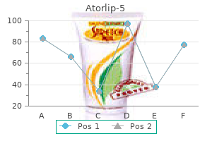
Atorlip-5 dosages: 5 mg
Atorlip-5 packs: 60 pills, 90 pills, 120 pills, 180 pills, 270 pills, 360 pills
Vascular provide Superior indirect receives its arterial supply instantly from the ophthalmic artery and indirectly from its supraorbital branch. Inferior rectus Inferior rectus arises from the widespread tendinous ring, beneath the optic canal. Innervation Superior indirect is innervated by the trochlear nerve, which enters the superior floor of the muscle. Actions Superior indirect is inserted into the posterior part of the eyeball; when it contracts, the again of the eyeball is elevated, and the front of the eyeball is depressed (particularly in the adducted position). Vascular supply Inferior rectus receives its arterial provide from the ophthalmic artery and from the infraorbital branch of the maxillary artery. Innervation Inferior rectus is innervated by a branch of the inferior division of the oculomotor nerve that enters the superior surface of the muscle. To acquire downward movement alone, inferior rectus should function with superior oblique. A fibrous extension from inferior rectus to the inferior tarsal plate of the eyelid causes the decrease eyelid to be depressed when the muscle contracts. Inferior indirect is a thin, slender muscle that lies close to the anterior margin of the floor of the orbit. It arises from the orbital surface of the maxilla lateral to the nasolacrimal fossa and ascends posterolaterally, at first between inferior rectus and the orbital flooring, and then between the eyeball and lateral rectus. The muscle broadens and thins, and, in contrast to the opposite extraocular muscular tissues, its tendon is barely discernible at its scleral attachment. Vascular supply Inferior oblique receives its arterial supply from the ophthalmic artery and from the infraorbital branch of the maxillary artery. Medial rectus Medial rectus is barely shorter than the opposite recti but is the strongest of the group. Innervation Inferior oblique is innervated by a branch of the inferior division of the oculomotor nerve that enters the orbital surface of the muscle. Actions Inferior oblique is inserted into the posterior part of the eyeball; when it contracts, the again of the eyeball is depressed and the front of the eyeball is elevated (particularly within the adducted position).
The human face is a fancy three-dimensional (3D) structure and capturing an image in its entirety with true-to-life precision is more likely to be difficult for both human and machine. A systematic review of methods of facial aesthetic evaluation in a particular patient population and a score system for end result comparability have been described beforehand (Sharma et al 2012). Traditionally, direct facial measurements have been taken using manual anthropometry utilizing rulers, tape measures and callipers, as pioneered by Leslie Farkas, the father of recent craniofacial anthropometry (Farkas 1994). Although low-cost and easy, this method requires a high degree of cooperation from the topic and could be very labour-intensive if used to assess lots of of individuals to acquire representative normative information. Digital images, against this, offers a rapid and everlasting picture that might be quickly processed and archived on to laptop databases. Drawbacks of utilizing a twodimensional (2D) modality to derive correct facial measurements embody differential positioning of subjects despite a standardization protocol and perspective projection distortion, where a 3D object is unavoidably misrepresented by an tried projection on to a 2D aircraft. Three-dimensional photogrammetry is a software-driven approach using multiple digital cameras set at completely different angles to purchase the facial picture quickly, in little over a millisecond. Time-of-flight technology, which measures the time taken for emitted light from an illumination unit to attain an object and journey back to a detector, has been used by a range of scanners to make very exact measurements of distance. This expertise makes use of either optical shutter know-how or modulated gentle of varied wavelengths to create a 3D picture of the actual face and head (Zhang and Lu 2013). However, there are currently no medical validation studies for facial evaluation using time-of-flight 3D cameras. Gross anatomical landmarks on the face are captured by a collection of relatively easy measurements. Delineation of finer options that may enable a detailed surface-based evaluation of facial form, nonetheless, requires a significantly larger number of densely arranged surface factors comparable to quasi-landmarks. A approach known as thinplate spline warping will then pull these factors on the surface of the face together, just like a thin sheet of rubber, into a exact alignment. The variety of landmarks chosen is a stability, as too few will cause the face scan to be registered poorly and too many will end in significant noise that causes the location of soft-tissue landmarks on a digital image to be inaccurate (Hammond et al 2004).
Diseases
There are four principal varieties, named filiform, fungiform, foliate and circumvallate papillae, and all besides the filiform papillae bear taste buds. Foliate papillae Foliate papillae lie bilaterally in two zones on the sides of the tongue close to the sulcus terminalis, each fashioned by a collection of pink, leaflike mucosal ridges, lined by a nonkeratinized epithelium. Their Circumvallate papillae Circumvallate papillae are giant cylindrical buildings, various in quantity from 8 to 12, which kind a Vshaped row immediately in front of the sulcus terminalis on the dorsal surface of the tongue. Several muscle tissue, including genioglossus and the posterior transverse (T) and vertical (V) intrinsic muscles, have been removed in order to observe individual nerves. The lateral branch is derived from the main trunk of the hypoglossal nerve as both a brief single branch (left facet in A) or multiple branches (right aspect in A and either side in B). Numerous style buds are scattered in both walls of the sulcus, and small serous glands (of von Ebner) open into the sulcal base. Numerous taste buds (pale buildings on the inner wall of the cleft, left side) are contained throughout the stratified epithelium of the papillary wall. They are quite a few on all kinds of lingual papillae (except filiform papillae), significantly on their lateral elements. Each style bud is linked by synapses at its base to certainly one of three cranial nerves that carry style, i. There is considerable individual variation in the distribution of taste buds in people. Taste buds have been described on the fetal epiglottis and soft palate but most disappear from these sites during postnatal development. Microstructure of taste buds Each style bud is a barrelshaped cluster of 50�150 fusiform cells, which lies within an oval cavity within the epithelium and converges apically on a gustatory pore, a 2 �m wide opening on the mucosal floor. The whole structure is about 70 �m in height by forty �m throughout and is sepa rated by a basal lamina from the underlying lamina propria.
At its perimeter, it has a gel-like consistency (100�300 �m thick); nearer the centre, it accommodates a more liquid zone. Hyaluronan, within the type of lengthy glycosaminoglycan chains, fills the whole vitreous. The cortex additionally contains scattered cells, the hyalocytes, which possess the characteristics of mononuclear phagocytes and should contribute to the manufacturing of hyaluronan. The liquid vitreous is absent at delivery, appears first at 4 or 5 years, and will increase to occupy half the vitreous house by the seventh decade. Vitreous liquefaction ends in an increased incidence of posterior vitreous detachment and related floaters in the elderly. The cortex is most dense at the pars plana of the ciliary physique adjacent to the ora serrata, where attachment is strongest, and this is often referred to as the base of the vitreous. Apart from the vitreous base, the vitreous additionally has a agency (peripapillary) attachment at the fringe of the optic disc. This adherence of the vitreous to the retina can result in traction on the retina if the vitreous shrinks, such as occurs in old age, resulting in macular holes or peripheral breaks, presumably leading to retinal detachment. In the fetus, this incorporates the hyaloid artery, which normally disappears about 6 weeks before birth. The canal persists in grownup life as a very delicate fibrous construction and is of no useful significance. Embryologically, the retina is derived from the 2 layers of the invaginated optic vesicle. The outer layer turns into a stratum of cuboidal pigment cells that separates the choroid from the neural retina, and subsequently varieties the outermost layer of the retina: the retinal pigment epithelium (layer 1). The different 9 strata of the retina develop from the internal layer of the optic vesicle and kind the neural retina. The outermost layer of the neural retina accommodates the light-sensitive elements of the photoreceptors, which convert the optical image into neural activity.
Anterior ethmoidal nerve the anterior ethmoidal nerve passes through the anterior ethmoidal foramen and canal, and enters the cranial cavity. It runs forwards in a groove on the upper floor of the cribriform plate beneath the dura mater, and descends via a slit lateral to the crista galli into the nasal cavity, the place it occupies a groove on the interior surface of the nasal bone and provides off the medial and lateral inner nasal branches. For an outline of the following distribution of the anterior ethmoidal nerve, see page 564. Ciliary ganglion the ciliary ganglion is a parasympathetic ganglion concerned with the innervation of sure intraocular muscle tissue. It is a small, flat, reddishgrey swelling, 1�2 mm in diameter, linked to the nasociliary nerve, and situated near the apex of the orbit in loose fat roughly 1 cm in entrance of the medial end of the superior orbital fissure. It lies between the optic nerve and lateral rectus, usually lateral to the ophthalmic artery. Its neurones, that are multipolar, are bigger than these found in typical autonomic ganglia; a really small variety of extra typical neurones are additionally current. Eight to ten delicate filaments, termed the short ciliary nerves, emerge anteriorly from the ganglion, arranged in two or three bundles, the decrease being larger. They run forwards sinuously with the ciliary arteries, above and under the optic nerve, and divide into 15�20 branches that pierce the sclera across the optic nerve and run in small grooves on the inner scleral surface. They convey parasympathetic, sympathetic and sensory fibres between the eyeball and the ciliary ganglion; solely the parasympathetic fibres synapse in the ganglion. The parasympathetic root, derived from the department of the oculomotor nerve to the inferior indirect, consists of preganglionic fibres from the Edinger�Westphal nucleus, which relay within the ganglion. Postganglionic fibres journey in the quick ciliary nerves to the sphincter pupillae and ciliary muscle. More than 95% of those fibres supply the ciliary muscle, which is way the bigger muscle in quantity. The sympathetic root contains fibres from the plexus around the inner carotid artery inside the cavernous sinus. These postganglionic fibres, derived from the superior cervical ganglion, form a fine branch that enters the orbit through the superior orbital fissure contained in the frequent tendinous ring. The fibres could cross directly to the ganglion, or may be part of the nasociliary nerve and journey to the ganglion in its sensory root; either method, they traverse the ganglion with out synapsing Posterior ethmoidal nerve the posterior ethmoidal nerve leaves the orbit by the posterior ethmoidal foramen and provides the ethmoidal and sphenoidal sinuses. Infratrochlear nerve the infratrochlear nerve leaves the orbit below the trochlea and supplies the skin of the eyelids, the conjunctiva, lacrimal sac, lacrimal caruncle and the facet of the nose above the medial canthus.
Syndromes
A newer classification system, aided by improved computed tomographic imaging, stresses the importance of the posterior ligamentous complex in figuring out the steadiness of the injured backbone along with the morphology of the damage (compression, translational/rotational or distraction injuries) and the neurological status of the patient (intact, nerve root injury, full or incomplete spinal twine injury, cauda equina syndrome) (Lee et al 2005). The elastic deformability of intervertebral discs permits tilting and axial rotation between vertebral our bodies, and likewise helps to scale back vertical accelerations of the pinnacle. The primary shock-absorbing mechanism of the column stems from the spinal curves, which enhance and reduce barely during locomotion in opposition to the restraining rigidity of the trunk muscle tissue. The elastic strain power in the stretched tendons of the muscle is responsible for shock absorption. These diurnal variations seem to be because of changes that happen within the cervical, thoracic and lumbar areas of the spine. The greatest change in vertebral column size is present in adolescents and younger adults. The top loss happens inside three hours of rising within the morning, with an total lack of about 15 mm. Although the curvatures inside the vertebral column contribute to the modifications in top, adjustments within the intervertebral disc contribute both to observed top loss and to variation in stability. Magnetic resonance imaging investigations reveal a dynamic movement of fluid into and out of an intervertebral disc and adjoining vertebral physique over a 24-hour period. In the early morning, the discs are swollen with water, the intervertebral ligaments and the anulus fibrosus are taut, and the intrinsic bending stiffness and stability of the osteoligamentous backbone are relatively excessive. After several hours of normal activity, the discs lose roughly 20% of their water and peak. This change makes the ligaments slack and greatly reduces the bending stiffness of the backbone, in order that comparatively more of the steadiness of the backbone should then be provided by the musculature. It is necessary to recognize the way in which by which the muscular tissues of the back work along side these of the abdominal wall, notably the indirect and transversus muscles, and with those of the lower limbs. The erector spinae group and internal oblique and transversus abdominis are anatomically and functionally linked by the thoracolumbar fascia (which encloses the previous, and into which the latter are inserted).
Activity of the medial efferents inhibits cochlear responses to sound; the strength of the activity grows slowly with growing sound stage. They are believed to modulate the micromechanics of the cochlea by altering the mechanical responses of the outer hair cells, thus altering their contribution to frequency selectivity and sensitivity. The firing rates of afferents from the left and right lateral canals are equivalent at relaxation (A). There are 30�40,000 nerve fibres within the human cochlear nerve (for evaluation, see Nadol (1988)). Their fibre diameter distribution is unimodal, and ranges from 1 to 11 �m, with a peak at 4�5 �m. Functionally, the nerve incorporates each afferent and efferent somatic fibres, together Autonomic nerve endings seem to be entirely sympathetic. Two adrenergic systems have been described inside the cochlea: a perivascular plexus derived from the stellate ganglion and a blood vessel-independent system derived from the superior cervical ganglion. Both methods travel with the afferent and efferent cochlear fibres and appear to be restricted to areas away from the organ of Corti. The sympathetic nervous system could trigger main and secondary effects within the cochlea by remotely altering the metabolism of assorted cell sorts and by influencing the blood vessels and nerve fibres with which it makes contact. Firing charges in the vestibular afferents that innervate receptors on either facet of the striola (red and green lines) are equivalent when the top is upright (A). When the pinnacle is tilted to the best (B) or to the left (C), the stereocilia are deflected by displaced otoconia; hair cells on the upward slope aspect of the striola enhance their firing rate, while these on the downward slope lower their firing rate. Peripheral auditory system Vibrations in the air column in the exterior acoustic meatus trigger a comparable set of vibrations within the tympanic membrane and auditory ossicles. The chain of ossicles acts as a lever that increases the force per unit area on the round window by 1.
The most superficial fibres prolong over three or four vertebrae, the deeper span two or three, and the deepest connect adjoining spines and are continuous with the interspinous ligament. Most of the ligament is fashioned by the tendons of muscles with posterior midline attachments, i. Their attachments extend from aspect joint capsules to the level the place laminae fuse to kind spines. Here their posterior margins meet and are partially united; the intervals between them admit veins that join the interior and posterior exterior vertebral venous plexuses. Their predominant tissue is yellow elastic tissue, whose nearly perpendicular fibres descend from the decrease anterior floor of 1 lamina to the posterior floor and upper margin of the lamina beneath. The anterior floor of the ligaments is roofed by a fantastic, continuous, easy lining membrane (Newell 1999). The ligaments are skinny, broad and long within the cervical area, thicker within the thoracic and thickest at lumbar ranges. For the detailed anatomy of the lumbar intertransverse ligaments, see Bogduk (2005). The transverse ligament is stronger than the dens, which therefore normally fractures before the ligament ruptures. The alar ligaments are weaker, and mixed head flexion and rotation could avulse one or each alar ligaments; rupture of 1 side ends in a rise of about one-third within the range of rotation to the opposite facet. Pathological softening of the transverse and adjoining ligaments or of the lateral atlanto-axial joints ends in atlanto-axial subluxation, which may trigger spinal wire injury. Laxity of cervical spinal ligaments may be a normal variant in children and result in diagnostic difficulties. Typical vertebral bodies are united by anterior and posterior longitudinal ligaments and by fibrocartilaginous intervertebral discs between sheets of hyaline cartilage (vertebral end-plates).
They anastomose with branches of the supraorbital, zygomatico orbital and lacrimal arteries. External (dorsal) nasal artery the exterior nasal artery is a termi nal department of the anterior ethmoidal artery, which arises from the ophthalmic artery. It emerges at the junction of the nasal bone and the lateral nasal cartilage and provides the skin overlaying the exterior nostril. Frontal (anterior) branch the frontal department passes upwards in direction of the frontal tuberosity and supplies the muscle tissue, pores and skin and peri cranium in this area. It anastomoses with its contralateral fellow and with the supraorbital and supratrochlear branches of the ophthalmic artery. Parietal (posterior) department the parietal branch is larger than the frontal department of the superficial temporal artery. It curves upwards and backwards, remains superficial to the temporal fascia, and anastomoses with its contralateral fellow and with the posterior auricular and occipi tal arteries. Accompanied by the larger occipital nerve, the occipi tal artery enters the back of the scalp by piercing the investing layer of deep cervical fascia that connects the cranial attachments of trapezius and sternocleidomastoid. Tortuous branches run between the pores and skin and the occipital belly of occipitofrontalis, anastomosing with the opposite occipital, posterior auricular and superficial temporal arteries, in addition to with the transverse cervical department of the subclavian artery. These branches provide the occipital belly of occipitofrontalis and the skin and pericranium associated with the scalp as far forwards because the vertex. Mental artery the psychological artery arises from the first a part of the maxil lary artery as a terminal branch of the inferior alveolar artery. It emerges on to the face from the mandibular canal at the psychological foramen, sup plies muscles and skin within the chin area, and anastomoses with the inferior labial and submental arteries. Posterior auricular artery the posterior auricular artery arises in the neck from the exterior carotid artery and ascends between the auricle and mastoid process. It provides 499 cHapTeR Supraorbital artery the supraorbital artery leaves the orbit through the supraorbital notch or foramen. It divides into superficial and deep branches that supply the skin and muscle of the higher eyelid, forehead and scalp. It anastomoses with the supratrochlear artery, frontal department of the superficial temporal and its contralateral fellow.
Traced laterally, the aponeurosis of the levator passes between the orbital and palpebral elements of the lacrimal gland to attach to the orbital tubercle of the zygomatic bone. Traced medially, it loses its tendinous nature as it passes closely over the reflected tendon of superior oblique, and continues on to the medial palpebral ligament as unfastened strands of connective tissue. Innervation Levator palpebrae superioris is innervated by a branch of the superior division of the oculomotor nerve that enters the inferior surface of the muscle. Sympathetic fibres to the graceful muscle part of levator palpebrae superioris (superior tarsal muscle) are derived from the plexus surrounding the inner carotid artery; these nerve fibres could be a part of the oculomotor nerve in the cavernous sinus and move forwards in its superior department. Superior rectus Levator palpebrae superioris Orbital apex Actions Levator palpebrae superioris elevates the upper eyelid. During this course of, the lateral and medial elements of its aponeurosis are stretched and thus restrict its action; the elevation can also be checked by the orbital septum. Levator palpebrae superioris is linked to superior rectus by a verify ligament; thus the upper eyelid elevates when the gaze of the attention is directed upwards. The place of the eyelids is dependent upon reciprocal tone in orbicularis oculi and levator palpebrae superioris, and on the degree of ocular protrusion. In the opened position, the higher eyelid covers the higher a half of the cornea, whereas the lower lid lies slightly below its lower margin. The eyes are closed by actions of both lids, produced by the contraction of the palpebral a part of orbicularis oculi and rest of levator palpebrae superioris. In wanting upwards, the levator contracts and the higher lid follows the ocular motion. At the identical time, the eyebrows are also usually raised by the frontal elements of occipitofrontalis to diminish their overhang. The lower lid lags behind ocular movement, in order that more sclera is uncovered beneath the cornea and the lid is bulged somewhat by the lower a part of the elevated eye. When the attention is depressed, each lids move; the higher retains its normal relation to the eyeball and nonetheless covers a few quarter of the cornea, whereas the decrease lid is depressed as a outcome of the extension of the thickened fascia of inferior rectus and inferior oblique pull on its tarsus as the former contracts. Extraocular muscle tissue Each rectus muscle passes forwards, within the place implied by its name, to be hooked up anteriorly by a tendinous growth into the sclera, posterior to the margin of the cornea. However, before their scleral attachment, the recti make functionally necessary connections inside orbital connective tissue that affect muscle action.
Mirzo, 63 years: If the excision line runs contrary to the skin pressure lines, the scar may be extra conspicuous and will tend to stretch transversely because of natural expressive facial movements. Tonsillectomy Surgical removing of the pharyngeal tonsils is often performed to prevent recurrent acute tonsillitis or to treat airway obstruction by hypertrophied or inflamed palatine tonsils.
Fasim, 23 years: A posterior pathway originates in the posterior eye area and passes to the brainstem saccadic mills through the superior colliculus. Hughes S, Yang H, ChanLing T 2000 Vascularization of the human fetal retina: roles of vasculogenesis and angiogenesis.
Fadi, 36 years: The mucosa of the nasopharynx is separated from the masticator house by the parapharyngeal space. B, A dorsal view of the developing vertebrae, with the centra within the middle and the bilateral parts of the neural processes laterally.
Farmon, 46 years: The superior laryngeal artery runs down towards the larynx, with the inner department of the superior laryngeal nerve mendacity above it. Lesions between C5 and T1 paralyse all four limbs (quadriplegia), the results in the upper limbs various with the positioning of injury: on the fifth cervical segment, paralysis is complete; and at the sixth, each arm is positioned in abduction and lateral rotation, with the elbow flexed and the forearm supinated, as a end result of unopposed exercise in deltoid, supraspinatus, rhomboid and the brachial flexors (all equipped by the fifth cervical spinal nerves).
Ramon, 47 years: The pores and skin over the junction of the bony and cartilaginous meatus is incised to permit the cartilage of the auricle and meatus to be swung forwards on its blood supply and so expose the bony meatus and mastoid course of. It con tains lymph nodes and small veins that unite to form the anterior jugular vein.
Mason, 32 years: The aim is to present increased and extra direct exposure of both the pathology and the adjacent important constructions with out the necessity to resect uninvolved structures. Hence, the nerves to the mandibular arch embrace the mandibular division of the trigeminal nerve, which is combined, and the chorda tympani and larger petrosal nerves, purely sensory pretrematic branches of the facial nerve.
References
Realice búsquedas en nuestra base de datos:
