
Jeffrey L. Anderson, MD, FACC, FAHA
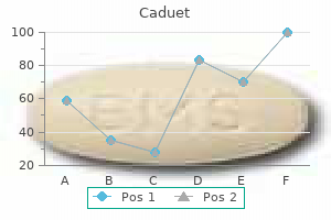
Caduet dosages: 5 mg
Caduet packs: 30 pills, 60 pills, 90 pills

First, enzyme-rich cortical granules within the oocyte cytoplasm release their contents (yellow) into the zona pellucida, beginning on the level of fusion and progressing right and left. Next, in the zona reaction, the enzymes modify the zona pellucida, transforming it into an impenetrable barrier to sperm as a guard towards polyspermy (multiple fertilization). Post-translational controls are sufficient and seem to contain regulation by way of protein phosphorylation. The means of compaction is essential for the technology of cell diversity in the early embryo. As every polarized cell divides, it retains significant components of its polar group, in order that its daughter cells inherit cytocortical domains, the nature of which displays their origin and organization within the original mother or father cell in the eight-celled embryo. Thus, if the axis of division is aligned approximately at proper angles to the axis of cell polarity, the more superficially placed daughter cell inherits all the apical cytocortex and some of the basolateral cytocortex and is polar, whereas the more centrally placed cell inherits solely basolateral cytocortex and is apolar. In distinction, if the axis of division is aligned approximately alongside the axis of the cell polarity, two polar daughter cells are fashioned. In this fashion, two-cell populations are fashioned within the 16-cell embryo that differ in phenotype (polar, apolar) and position (superficial, deep). The number of cells in each population in anybody embryo will be determined by the ratio of divisions along, and at proper angles to , the axis of eight-cell polarity. The theoretical and observed limits of the polar to apolar ratio are 16: zero and eight: eight. In cleavage, the generation of cell variety, to either trophectoderm or inner cell mass, happens in the 16-cell morula and precedes the formation of the blastocyst. During the 16-cell cycle, the outer polar cells proceed to differentiate an epithelial phenotype, and show additional features of polarity and intercellular adhesion typical of epithelial cells, while the internal apolar cells stay symmetrically organized. During the subsequent cell division (16�32 cells), a proportion of polar cells once more divide differentiatively, as within the earlier cycle, each yielding one polar and one apolar progeny, which enter the trophectoderm and internal cell mass lineages, respectively.
Syndromes
This weak, reversible adhesion allows leukocytes to roll along the endothelial floor of a vessel lumen at low velocity, making and breaking contact, and sampling the endothelial cell surfaces. Finally, the leukocyte migrates via the vessel wall (diapedesis), passing either between (paracellular migration) or throughout (transcellular migration) endothelial cells. Transcellular migration is assumed to be the popular pathway; endothelial transcytotic vesicles (caveolae), intermediate filaments (vimentin) and F-actin are necessary within the creation of transient transcellular channels through which leukocytes cross. There are three identified members of the selectin family of adhesive proteins: L-selectin (also often identified as lymphocyte homing receptor), E-selectin and P-selectin. L-selectin mediates homing of lymphocytes, particularly to peripheral lymph nodes, however also promotes the accumulation of neutrophils and monocytes at sites of inflammation. E-selectin is an inducible adhesion molecule that mediates adhesion of leukocytes to inflammatory cytokine-activated endothelium, and is simply transiently expressed on endothelium. It binds to ligands expressed on neutrophils, platelets and monocytes, and, like E-selectin, tethers leukocytes to endothelium at websites of irritation. P-selectin is shortly endocytosed by the endothelial cells and so its expression is short-lived. The integrins are a big family of molecules that mediate cell-to-cell adhesion as nicely as interactions of cells with extracellular matrix. In contrast to 1 integrins, which many cells specific, the expression of 2 integrins is restricted to white blood cells. Three members of the big immunoglobulin superfamily of proteins are concerned in leukocyte�endothelial adhesion, offering integrin counter-receptors on the endothelial cell membrane. Erythrocytes and leukocytes (mainly lymphocytes and neutrophils) are seen within the lumen. It accommodates a typical fibrocollagenous extracellular matrix, a number of fibroblasts and occasional easy muscle cells. Endothelial von Willebrand issue concentrates in this layer and participates in haemostasis and platelet adhesion when the overlying endothelium is broken.
Racial variations in colour are mainly as a end result of variations within the amount, type and distribution of melanin and are genetically decided. These genetic variants may also decide the prevalence of benign pores and skin lesions such as freckles, in addition to susceptibility to frequent types of non-melanoma skin cancers. The common state of well being is mirrored within the appearance and condition of the skin, and the earliest indicators of many systemic disorders may be obvious in the skin. Other cells within the dermis embody melanocytes (pigment-forming cells from the embryonic neural crest), Langerhans cells (immature antigen-presenting dendritic cells derived from bone marrow), lymphocytes and Merkel cells. Merkel cells may perform as sensory mechanoreceptors or presumably as a half of the dispersed neuroendocrine system and lots of are associated with nerve endings. In routine histological preparations, the nonkeratinocyte cells are almost indistinguishable, and appear as clear cells surrounded by a space produced by shrinkage throughout processing. This turnover of keratinocytes is mediated by stem cells that reside within the basal layer of the epidermis. These basal cells generate daughter cells, which endure a sequence of biochemical and physical changes as they migrate towards the surface of the pores and skin to kind the assorted layers of the epidermis. They remodel from polygonal dwelling cells to non-viable flattened squames filled with intermediate filament proteins (keratins) embedded in a dense matrix of cytoplasmic proteins to form mature keratin. The first three of those layers are metabolically lively compartments by way of which cells pass and change their morphology as they endure mobile differentiation. The epidermis has been partially peeled back to present the interdigitating dermal and epidermal papillae. The epidermal appendages (pilosebaceous models, sweat glands and nails) are shaped developmentally by the ingrowth of the dermis. The basal layer or stratum basale is the innermost layer of the dermis and is adjacent to the dermis.
The rectum is at all times affected and the proximal extent of the situation is variable. The condition can also be characterized by a transition zone with decreased innervation. It is important to remember that the transition zone could comply with an uneven course around the circumference of the bowel and a single biopsy may be unreliable. Of the neural crest cells that colonize the bowel, some within the foregut could purchase the flexibility to migrate outwards and colonize the developing pancreas. Chromaffin cells Chromaffin cells are derived from the neural crest and found at numerous websites all through the physique. They are the traditional chromaffin cells of the suprarenal medulla, bronchial neuroepithelial cells, dispersed epithelial endocrine cells of the gut (formerly often identified as argentaffin cells), carotid physique cells, and the paraganglia (Ch. The sympathetic ganglia, suprarenal medulla and chromaffin cells are all derived from the cells of the sympathosuprarenal lineage. In the suprarenal medulla, these cells differentiate into a selection of types consisting of small and intermediate-sized neuroblasts or sympathoblasts, and larger, initially rounded phaeochromocytoblasts. Large cells with pale nuclei, thought to be the progenitors of chromaffin cells, can be detected from 9 weeks in human fetuses, and clusters of small neurones are evident from 14 weeks. Intermediate-sized neuroblasts differentiate into the standard multipolar postganglionic sympathetic neurones (which secrete noradrenaline (norepinephrine) at their terminals) of classic autonomic neuroanatomy. The massive cells differentiate into masses of columnar or polyhedral phaeochromocytes (classic chromaffin cells), which secrete both adrenaline (epinephrine) or noradrenaline (norepinephrine). These cell masses are termed paraganglia and could also be situated near, on the surface of, or embedded in, the capsules of the ganglia of the sympathetic chain, or in some of the large autonomic plexuses. The largest members of the latter are the para-aortic our bodies that lie along the perimeters of the abdominal aorta in relation to the inferior mesenteric artery. During childhood, the para-aortic our bodies and the paraganglia of the sympathetic chain partly degenerate and might no longer be isolated by gross dissection, but even in the adult, chromaffin tissue can nonetheless be acknowledged microscopically in these varied sites. Enteric nervous system the enteric nervous system is completely different from the opposite elements of the autonomic nervous system because it could mediate reflex exercise independently of control by the mind and spinal wire. The neuroblasts of the lateral walls of the tube are giant and, at first, spherical or oval (apolar). Further differentiation leads to the development of dendritic processes and the cells turn out to be typical multipolar neurones.
A basic animal experiment concerning bone mechanobiology, which is still widely cited. Thambyah A, Broom N 2007 On how degeneration influences loadbearing in the cartilagebone system: a microstructural and micromechanical study. Wang H, Listrat A, Meunier B et al 2014 Apoptosis in capillary endothelial cells in ageing skeletal muscle. The muscle in these tubes is of two sorts: easy muscle is attribute of the partitions of blood vessels, whereas cardiac muscle offers the walls of the heart chambers with their powerful contractile pumping action. The basic characteristics and classification of muscle tissues are given on page 103. Smooth muscle also forms an important contractile factor in the partitions of many different organ techniques of the body. Smooth muscle can additionally be referred to as involuntary muscle as a result of its exercise is neither initiated nor monitored consciously. It is far more variable, in each form and performance, than both striated or cardiac muscle, a mirrored image of its diversified roles in different techniques of the body. Smooth muscle is typically found in the partitions of tubular buildings and hollow viscera. The account that follows will therefore be involved with the generic properties of clean muscle. The more specialized morphologies of smooth muscle are described in the acceptable regional chapters. Such an arrangement achieves both shut packing and a more efficient switch of drive from cell to cell. This look contrasts markedly with that of skeletal muscle cells, which present a constant diameter in cross-section and peripherally placed nuclei throughout their length. Smooth muscle has no attachment structures equal to the fasciae, tendons and aponeuroses associated with skeletal muscle. There is a particular arrangement for transmitting pressure from cell to cell and, the place essential, to other gentle tissue buildings. These components bridge the gaps between adjacent cells and provide mechanical continuity throughout the fascicle. At the boundaries of fascicles, the connective tissue fibres become interwoven with those of interfascicular septa, so that the contraction of various fascicles is communicated throughout the tissue and to neighbouring structures.
Beta-alanyl-L-histidine (Carnosine). Caduet.
Source: http://www.rxlist.com/script/main/art.asp?articlekey=96994
Brainstem the medial rectus subnucleus consists of three anatomically distinct subpopulations. The ventral portion, which contains the biggest variety of motor neurones, occupies the rostral two-thirds; a subpopulation of smaller motor neurones lies dorsally all through the rostral two-thirds of the nucleus and innervates the small orbital fibres of the medial rectus; and one other subpopulation lies dorsolaterally in the caudal twothirds of the nucleus. A median subnucleus of enormous neurones, the caudal central nucleus, lies at the caudal pole of the oculomotor nucleus adjacent to the superior rectus and medial rectus subnuclei. In non-human primates, roughly 30% of the motor neurones on this subnucleus innervate levator palpebrae superioris bilaterally, which is exclusive for paired skeletal muscle tissue. Its neurones give rise to axons that journey within the oculomotor nerve and synapse with postganglionic neurones in the ciliary ganglion (Kozicz et al 2011). Fascicles of axons from these subnuclei course forwards within the midbrain and emerge because the oculomotor nerve in the interpeduncular fossa. The fascicles are most probably organized from medial to lateral subserving the pupil, inferior rectus, medial rectus, levator palpebrae superioris and superior rectus, and inferior oblique. Afferent inputs to the oculomotor nuclear complicated include fibres from the rostral interstitial nucleus of the medial longitudinal fasciculus and the interstitial nucleus of Cajal, each of which are concerned in the control of vertical and torsional gaze. Other inputs come, both immediately or indirectly, from the nuclei of the posterior commissure, the interstitial nucleus of Cajal, the frontal eye fields, the superior colliculus, the dentate nucleus and other cortical areas. The medial longitudinal fasciculus carries connections from the trochlear, abducens and vestibular nuclei; the medial and lateral vestibular nuclei project to the medial rectus subnucleus. Afferent inputs to the Edinger�Westphal preganglionic nucleus come primarily from the pretectal nuclei bilaterally, mediating the pupillary gentle reflex, and from the visual cortex, mediating accommodation. Most efferent fibres from the inferior colliculus journey through the inferior brachium to the ipsilateral medial geniculate nucleus. Lemniscal fibres relay solely within the central nucleus, and some pass with out relay to the medial geniculate nucleus. In humans, the ventral division of the medial geniculate nucleus receives a topographic projection from the central nucleus and the dorsal division receives an analogous projection from the dorsal cortex. A descending projection from the auditory cortex reaches the inferior colliculus via the medial geniculate nucleus.
Spinomesencephalic fibres are mostly myelinated and ascend in the white matter of the ventrolateral quadrant of the spinal wire, in association with the spinothalamic and spinoreticular tracts. Spinomesencephalic neurones are of low-threshold, wide-dynamicrange or high-threshold lessons. Their receptive fields could additionally be small, or very advanced and embody giant surface areas of the physique. Many spinomesencephalic cells are nociceptive and are likely to be concerned within the motivational�affective part of pain. Electrical stimulation of their site of termination within the periaqueductal gray matter leads to severe pain in humans. Furthermore, the cells of the deeper layers of the superior colliculus, where spinotectal fibres synapse, are activated by noxious stimuli. Axons forming the tract cross and then ascend superficially on the junction of the ventral and lateral white funiculi, to finish within the dorsal and medial accessory olivary nuclei. The tract carries data from muscle and tendon proprioceptors, and likewise from cutaneous receptors. A functionally comparable route, the dorsal spino-olivary tract, ascends within the dorsal white funiculi, and relays within the dorsal column nuclei to the contralateral inferior olivary nucleus. Information on these tracts in primates is scant, but postmortem evidence following cordotomies in people has revealed degenerating axonal terminals within the inferior olivary nucleus. Evidence from animal studies means that cells of origin occur at all levels of the spinal cord, notably within the upper cervical segments. The sample of anterograde degeneration, in each human postmortem research and in experimental animals following anterolateral cordotomy, signifies the existence of spinoreticular projections to many nuclei of the medial pontomedullary reticular formation. There is also a projection to the lateral reticular nucleus (a precerebellar relay nucleus). Innocuous cutaneous stimuli may inhibit or excite a particular cell, whereas noxious stimuli are often excitatory. They are concerned with the control of movement, muscle tone and posture, the modulation of spinal reflex mechanisms and the modulation of transmission of afferent data to higher ranges. Corticospinal and corticonuclear tracts Corticospinal and corticonuclear fibres come up from neurones within the cerebral cortex. The majority of corticospinal and corticonuclear fibres arise from cells located within the main motor cortex (area 4) and the premotor cortex (area 6).
The abducens nerve emerges on the caudal border of the pons generally according to the rootlets of the hypoglossal nerve. Caudally, most fibres of the pyramid taper, decussate and enter the lateral funiculus of the spinal twine. The posterolateral sulcus is lateral to the olive; the glossopharyngeal and vagus nerves be part of the brainstem along the line of this sulcus, according to the dorsal spinal nerve roots. The spinal central canal extends into the caudal half of the medulla, migrating progressively extra dorsally till it opens out into the lumen of the fourth ventricle on the obex. In the closed a part of the medulla, a shallow dorsal intermediate sulcus, on either side of the dorsal median sulcus, is continuous with its cervical spinal counterpart and indicates the location of the dorsal (posterior) columns (fasciculi gracilis and cuneatus). The restiform body lies dorsolaterally in the medulla, forming a rounded ridge between the caudal a half of the fourth ventricle and the glossopharyngeal and vagal rootlets on each side. The restiform our bodies on the two sides diverge and incline to enter the cerebellar hemispheres as the main part of the inferior cerebellar peduncles. Key: 1, infundibulum; 2, tuber cinereum; three, mammillary body; 4, basilar pons; 5, abducens nerve; 6, foramen caecum; 7, olive; eight, glossopharyngeal nerve; 9, vagus nerve; 10, rootlets of hypoglossal nerve; eleven, accessory nerve; 12, olfactory tract; thirteen, optic nerve; 14, optic chiasma; 15, optic tract; sixteen, oculomotor nerve; 17, uncus; 18, trochlear nerve; 19, trigeminal nerve; 20, facial nerve; 21, vestibulocochlear nerve; 22, flocculus; 23, pyramid; 24, motor decussation (decussation of pyramids). The inferior cerebellar peduncles kind the anterior and rostral boundaries of the lateral recesses of the fourth ventricle; these are continuous with the subarachnoid house through the lateral apertures of the fourth ventricle (foramina of Luschka). A tuft of choroid plexus, continuous with that of the fourth ventricle, protrudes from the foramina on either side. The decussation displaces the central grey matter and central canal dorsally (Haines 2015). Continuity between the ventral gray column and central grey matter, which is maintained throughout the spinal twine, is misplaced. The column subdivides into the supraspinal nucleus (continuous above with that of the hypoglossal nerve), which is the efferent source of the primary cervical nerve, and the nucleus of the accent nerve, which is in line rostrally with the nucleus ambiguus. The flooring of the fourth ventricle has been exposed by cutting the cerebellar peduncles and removing the cerebellum.
In a large limb muscle, a motor neurone may internal vate several hundred muscle fibres. Within a muscle, fibres belonging to one motor unit are distributed over a wide territory, without regard to fascicular boundaries, and so they intermingle with the fibres of other motor units. Motor units become larger in cases of nerve injury as a result of denervation induces collateral or terminal sprouting of the remaining axons. Each new branch can reinnervate a fibre, thus increas ing the territory of its father or mother motor neurone. Excitation�contraction coupling is the method whereby an motion potential triggers the release of calcium from the terminal cister nae of the sarcoplasmic reticulum into the cytosol. This activates a calciumsensitive swap within the thin filaments and so initiates contrac tion. As the overlap increases, the I and Hbands slim to nearextinction, while the width of the Abands remains fixed. Filament sliding is dependent upon the making and breaking of bonds (crossbridge cycling) between myosin head regions and actin filaments. Actin filament binding sites for myosin are revealed solely by the presence of calcium, which is released into the sarcoplasm from the sarcoplasmic reticulum, causing a repositioning of the troponin�tropomyosin advanced on actin: this is the calciumsensitive change. Slow-twitch versus fast-twitch fibres the passage of a single action potential by way of a motor unit elicits a twitch contraction where peak pressure is reached within 25�100 ms, relying on the motor unit sort involved. However, the motor neurone can ship a second nervous impulse in much less time than it takes for the muscle fibres to relax. When this happens, the muscle fibres contract again, constructing the stress to a higher degree. In practice, the 2 mecha nisms seem to operate in parallel, however their relative importance may rely upon the dimensions and/or function of the muscle; in massive muscles with many motor models, motor unit recruitment might be the more impor tant mechanism.
The sample of limb growth is managed by data contained within the somatopleuric mesenchyme. Regions of the limb are specified by interplay between the floor ectoderm (apical ectodermal ridge) and underlying somatopleuric mesenchyme; together, these tissues type the progress zone of the limb. The somato pleuric mesenchyme within the limb bud additionally specifies the postaxial border of the growing limb. Somatopleuric mesenchyme offers rise to the connective tissue parts of the appendicular skeleton, including the pectoral and pelvic girdles and the bones and cartilage of the limbs, and their associated ligaments and tendons. It additionally offers rise to the dermis of the pores and skin of the ventral and lateral body walls and of the limbs. The individual tissue elements have been separated but are aligned in register through the numbered zones. It performs a patterning function in endodermal devel opment, specifying the region and villus sort in the gut, and the department ing sample within the respiratory tract. The mesenchyme cells are arranged as layers, one steady with the dorsal side of the par axial mesenchyme and the somatopleure, the opposite with the ventral aspect of the paraxial mesenchyme and the splanchnopleure. As growth proceeds, the intermediate mesenchyme varieties a loosely packed dorsolateral wire of cells, which lengthens on the caudal end and in the end joins the cloaca. Angioblastic mesenchyme Mesenchymal cells, which give rise to the cellular parts of the blood, the endothelium and the mesenchymal layers of the tunica externa and adventitia of blood and lymphatic vessels, arise from extraembryonic and intraembryonic sources. Evidence suggests that endodermal tissues are needed for endothelial differentiation, par ticularly the early foregut. Angioblastic mesenchyme varieties early within the third week of improvement from extraembryonic mesenchyme within the splanchnopleure of the yolk sac, in the body stalk (containing the allantois), and in the somatopleure of the chorion. The peripheral cells flatten as a vascular endothelium, whereas the central cells remodel into primitive pink blood corpuscles.
Kapotth, 27 years: It projects to the supplementary motor area, the dorsal premotor cortex and the frontal eye area. First, the midbrain harm entails constructions on the facet of the herniation with predictable deficits: ipsilateral pupil dilation and ophthalmoplegia, contralateral weak point of upper and lower extremities, an altered level of consciousness and hyperactive reflexes. The mastoid course of appears within the second 12 months and the metopic suture between the two frontal bones is usually closed by the end of the primary 12 months. In the dorsal column nuclei, the lower limb is represented within the nucleus gracilis, the upper limb within the nucleus cuneatus, and the trunk in an intermediate place between them.
Lee, 34 years: These may be primarily cutaneous vessels, which provide the pores and skin immediately but contrib ute small branches to the muscle as they cross via it, or they may be the terminal branches of intramuscular vessels, which go away the muscle to complement the cutaneous blood provide. This main distinction between cardiac and skeletal muscle is mirrored in tissue-specific isoforms of calcium release channels. During the formation of trophoblast, hypoblast (primitive endoderm) and epiblast, the cells reply regionally to their setting, particularly to other cells that are positioned lateral and basal to them. The dorsal ramus additionally gives off a spinal department, which enters the vertebral canal and divides into a sequence of branches that supply the partitions and joints of the osteoligamentous canal, and neural branches to the spinal twine and spinal nerve roots.
Tjalf, 44 years: This paper presents an extensive profile of the normal human amniotic fluid proteome and considers the molecular features of amniotic fluid including the development of biomarkers for pregnancy-associated abnormalities. Holland A, Ohlendieck K 2013 Proteomic profiling of the contractile appa ratus from skeletal muscle. As layers of calcifying matrix are added to the early trabeculae, osteoblasts become enclosed inside primitive lacunae. The vestibular nuclei project extensively to the cerebellum and in addition receive axons from the cerebellar cortex and the fastigial nuclei.
References
Realice búsquedas en nuestra base de datos:
