

Javier Bolanos Meade, M.D.

https://www.hopkinsmedicine.org/profiles/results/directory/profile/0017464/francisco-bolanos-meade
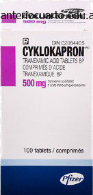
Cyklokapron dosages: 500 mg
Cyklokapron packs: 30 pills, 60 pills, 90 pills, 120 pills, 180 pills, 270 pills
These perforations provide avenues of egress for accumulated subgraft fluid in a lot the same way that meshing does for split-thickness grafts. Allow approximately 7 minutes for the epinephrine to take effect and help reduce bleeding throughout harvest. The full-thickness pores and skin graft can be stretched over the finger and curved scissors used to instantly excise fats from the undersurface of the full-thickness graft. The removing of unwanted fats maximizes the surface space of deep dermis in direct contact with the wound bed, which helps to facilitate the inosculation and revascularization process. Graft Placement To enhance the precision of dermal edge contact of the graft in the wound, suture fixation is most well-liked over staple fixation. The process for dressing this graft is similar as that for a split-thickness graft: using either Aquaphor or Xeroform gauze dressings with an overlying bolster of Reston foam or cotton batting secured with gauze and an elastic wrap, followed by appropriate immobilization of the realm. Graft Preparation To decrease the accumulation of hematoma or seroma beneath the graft, a well-prepared mattress is required. They turn into fibrovascularly built-in into the wound mattress as a synthetic neodermis. The technical utility of AlloDerm and Integra is the same as placement of a full-thickness pores and skin graft. AlloDerm Biobrane When utilizing AlloDerm, bolstering dressings are applied over a petrolatum-doped nonadherent gauze interface, and the dermal assemble is observed periodically at twice-weekly intervals. AlloDerm will reveal granulation tissue issuing by way of the pores of the dermal assemble, typically at about 2 to three weeks after graft placement. Integra On preliminary placement, Integra appears white, with a transition over the succeeding 2 to three weeks to a rosy colour, the byproduct of neovascularization. At this level the Silastic layer of the Integra can be separated and the vascularized dermal assemble grafted with a thin splitthickness pores and skin graft.
A direct posterior incision has been criticized for an elevated propensity towards postoperative seroma formation. This patient developed stiffness after nonoperative treatment of a nondisplaced radial neck fracture. Advantages to the lateral exposure embody its simplicity, less extensor and flexor�pronator disruption, and entry to all three joint articulations. The main disadvantage of the lateral publicity is the inability to handle the ulnar nerve when indicated. For the posterior incision, care is taken to keep away from putting the road of incision directly over the prominence of the olecranon. Full-thickness fasciocutaneous flaps are elevated laterally to expose the extensor muscle mass. A triceps tenolysis is carried out with an elevator, releasing any adhesions between the muscle and the posterior humerus. The humeroulnar joint is recognized posteriorly and the olecranon fossa is cleared of any fibrous tissue or scar that may limit terminal extension. The anconeus and triceps are reflected posteriorly, exposing the posterior capsule, olecranon tip, and olecranon fossa. Visualization of the posterior compartment permits d�bridement of the posterior joint, together with eradicating impinging tissue of osteophytes in the olecranon fossa and the tip of the olecranon. The proximal edge of this complex lies along the proximal border of the radial head. Dissection is then carried out beneath the elbow capsule between the joint and the brachialis. The radial and coronoid fossae are cleared of fibrous tissue and the tip of the coronoid is removed if overgrowth or impingement was famous in flexion. After launch of the anterior capsule, gentle extension of the elbow with utilized pressure usually brings the joint out to practically full extension. In longstanding circumstances of contracture, the brachialis muscle could be tight, inhibiting full terminal elbow extension.
We only include fractures with three or fewer articular fragments, which meet criteria for fractures that can be operatively decreased and stuck with reproducibly good outcomes. The radiocapitellar joint has a saddle-shaped articulation permitting each flexion and extension in addition to rotation. The radial head, a secondary stabilizer, maintains up to 30% of valgus resistance within the native elbow. It may be prudent to protect a repaired radial head from excessive valgus stress throughout early range of movement by inserting a hinged exterior fixator. The radial head additionally features in the transmission of axial load, transmitting 60% of the load from the wrist to the elbow. A fall on an outstretched hand with the elbow in extension and the forearm in pronation produces an axial or valgus load (or both) driving the radial head into the capitellum, fracturing the relatively osteopenic radial head. The radial recurrent artery, a department of the radial artery, provides the main blood supply to the radial head. In most cadaveric specimens, a branch of the radial recurrent penetrates the radial head in the secure zone. Radial head resection should be reserved for patients with low useful demands or restricted life expectancy, and when the surgeon has excluded elbow instability with a fluoroscopic examination. Delayed excision of the radial head after failed nonoperative management could additionally be thought of with modest improve in function; it has proven 23% honest or poor outcomes at 15 years of follow-up. Range-of-motion and stress examinations are important to proper choice making and may obviate the necessity for advanced imaging if carried out accurately with adequate anesthesia. If omitted, this can lead to undiagnosed associated injuries and should lead to flawed determination making. In the emergency department or workplace, enough anesthesia may be obtained by aspirating hematoma, then injecting the elbow joint with 5 mL of local anesthetic and inspecting the elbow beneath fluoroscopy. Normal values are zero to a hundred forty five degrees of flexion�extension, eighty five degrees of supination, and eighty levels of pronation. A radiocapitellar view with forearm in neutral and at 45 levels cephalad gives an improved view of the articular surfaces.
Arterialization of the venous system for the treatment of end-stage ischemia of the higher extremity. The commonest infecting organism is Staphylococcus aureus, though these infections are generally combined infections. A chronic paronychia is characterized by repeated infection and irritation of the eponychium. This problem often occurs within the setting of repeated and prolonged exposure to water. The most commonly isolated organisms are Candida albicans, gram-positive cocci, gram-negative rods, and Mycobacterium spp. Herpetic whitlow is caused by an outbreak of herpes simplex virus within the pores and skin of the finger and may be confused with acute paronychia or felon. This commonly occurs on account of a hangnail, nail biting, or an overzealous manicure. Chronic paronychia results from colonization and infection by organisms that enter the space between the nail plate and the cuticle, eponychium, and nail fold. This chronic an infection and inflammation lead to fibrosis of the eponychium, which, in flip, leads to decreased vascularity of the dorsal nail fold. Felons typically outcome from penetrating trauma, or from bacterial inoculation through the exocrine sweat glands contained inside the pulp. Cellulitis and native irritation lead to local ischemia, which, in the setting of the closed spaces defined by septa, leads to increased pressure. Fat necrosis and abscess formation end result from the increased strain, which, in turn, causes an additional enhance in stress, and, in effect, a compartment syndrome. The eponychium is the tissue that attaches carefully to the nail plate proximally, commonly referred to as the cuticle.
After removing this graft, harvest cancellous bone from the location and tightly pack it between the ready bony surfaces. In circumstances of extreme deformity, the carpus may be held generally alignment with temporary Kirschner wires. Key the corticocancellous graft into the space between the third metacarpal base and the radius platform. Choose the specified wrist fusion plate and secure it distally to the third metacarpal with appropriately sized screws. In chosen situations, the second metacarpal may be used rather than the third metacarpal. If needed, the extensor retinaculum could also be split, with one portion repaired deep to the extensor tendons to enable coverage of distinguished parts of the plate. Joints throughout the wrist which would possibly be decorticated and grafted: elective (O) or required (R). The graft is keyed into the area between the third metacarpal base and the radius platform. Typically, cancellous autograft taken from the distal radius is used between the prepared bony surfaces. These are usually smaller pins that produce an interference fit within the radius shaft. Choose the most important pin that will fit throughout the metacarpal and advance it retrograde by way of the lowered carpus and into the radius. Complex wrist collapse secondary to rheumatoid arthritis handled with an intramedullary rod and wiring. Alternatively, a determine eight wire may be positioned across the third metacarpal and thru the radius to compress the assemble. If the metacarpophalangeal joints have already been changed, two Steinmann pins via the second and third internet areas may be effective. Patients choose to be in slight wrist extension without significant radial or ulnar deviation; important deviation into flexion or radial deviation results in problems and weak spot. Patients with an extensor lag as a outcome of dorsal swelling are started on a program of dynamic extension with an outrigger splint until full lively extension is regained.
Syndromes
The myotendinous junction for the wrist extensors is in the mid-forearm, whereas the myotendinous junction of the finger and wrist extensors is the distal forearm. Multiple triceps motor branches are current because the nerve programs within the posterior compartment of the upper arm. The nerve then lies between the brachialis and brachioradialis earlier than it enters the forearm. The motor level for each nerve is pretty consistently positioned just proximal to the myotendinous junction. The sequence of muscle innervation is a crucial distinction when contemplating the anatomy of the radial nerve. The order of innervation is essential in differentiating a radial nerve harm from a mechanical myotendinous harm or muscle disruption after a forearm laceration. Understanding the innervation also is helpful while observing and assessing the scientific restoration after radial nerve injury or restore. Radial nerve damage is most commonly associated with midto distal shaft humerus fractures. The aim of therapy is independent wrist, finger, and thumb extension with thumb abduction. Conventional surgical recommendations are to proceed after the patient has reached a documented medical and electromyographic plateau of useful radial nerve regeneration. The medical recovery may be adopted by observing the advancing Tinel sign and the reinnervation sequence. In the medical setting of a nonadvancing Tinel sign and electromyographic findings of axonal loss, exploration with intraoperative electrophysiologic testing is warranted. The brachioradialis could be palpated throughout resisted, neutral position elbow flexion, and the wrist assumes a radial deviated position during attempted active extension. In common, the optimal rigidity is established at the peak of the length�tension curve for the donor muscle, whereas the wrist and fingers are maintained in the perfect position. Because this donor muscle position is difficult to determine intraoperatively without specialised equipment, this point moderately corresponds to the midpoint of the passive muscle tour. The best joint place for each transfer is mentioned with the person transfers.
The prognosis is extraordinarily poor with these tumors, with survival instances averaging 2. In this patient population, the illness has an indolent course and may be treatable with surgical procedure and radiation. Hemangiopericytoma is a diffuse proliferation of capillaries, encased in connective tissue and surrounded by pericytes. They might current as a nonpigmented bleeding mole, an ulceration with outstanding telangiectasia, or a darkish blue, hemorrhagic swelling. The affected person offered with a big mass of the forearm that had been present for forty six years. This may help to differentiate between a hemangioma and an arteriovenous malformation in early childhood. The physician should ascertain whether or not there are any compressive symptoms from the lesion according to a mass effect, distention, or ache with train that might point out a venous malformation. It is also important to ask about any signs of congestive coronary heart failure, which can happen as sequelae of an untreated high-flow malformation. Glomus tumors the classic triad of paroxysmal ache, pinpoint tenderness, and temperature intolerance, especially cold, must be elicited if glomus tumors are in the differential. On bodily examination, the doctor ought to search for a bluish discoloration (found in 28% of patients) and a pulp nodule or nail deformity (found in 33% of patients). This might help to decide whether or not the lesion is an incomplete excision or a new tumor. If colour returns to the radial aspect of the hand inside 5 seconds, the superficial arch is complete and the radial artery may be ligated. Methods for analyzing the vascular lesions of the hand the examiner should look at the hand to check for blue spots, nail ridging, reddish, raised lesions, pulsatile lots, or traumatic damage, which helps to differentiate between malformations, aneurysms, pyogenic granulomas, and glomus tumors. In fast-flow arteriovenous malformations, a bruit or thrill may be heard, which might not be found in other vascular lesions. If the patient has diminished or resolved ache with this maneuver, then the take a look at is considered positive for a glomus tumor. It can be utilized to outline the anatomic extent of lesions and their relationship with the encircling tissue.
Classification Although a number of classification techniques have been proposed, the most commonly used is the Mayo Radiographic Classification System (Table 1). The grading system relies on bone high quality, joint area, and bony structure and delineates 4 grades of progression so as of increasing severity. Judicious use of intra-articular steroid injections additionally performs a job in symptom administration. Aggressive administration of the synovitis can restrict or delay the onset and severity of joint involvement. Physical Therapy the objective of bodily therapy is to encourage range of movement, useful power, and upkeep of activities of day by day living. The major goal of nonoperative administration of the rheumatoid elbow is to minimize delicate tissue swelling and For early illness states, wonderful scientific outcomes may be achieved with synovectomy carried out utilizing open or arthroscopic strategies. Although this process has not essentially been shown to alter the pure history of the illness, it reliably produces symptomatic aid for five or extra years in the majority of circumstances performed on elbows within the early levels of the illness process. When open synovectomy is carried out, the radial head must be excised to entry and completely d�bride the diseased synovial tissue that exists in this region. Open synovectomy has traditionally been accompanied by radial head excision due to (1) ubiquitous radiocapitellar and proximal radioulnar joint articular destruction and (2) the necessity to surgically expose the sacciform recess for the requisite full synovectomy. Otherwise, a complete arthroscopic synovectomy is performed without excising the radial head. In addition, the minimally invasive nature of an arthroscopic approach yields the potential benefits of less ache, sooner recovery with earlier range of movement, and a lower fee of infection in contrast with an open process. An arthroscopic anterior capsular launch may be performed at the time of the arthroscopic synovectomy to enhance elbow extension. A posterior olecranon-plasty may be carried out to re-establish regular concavity of the olecranon fossa. Posteromedial capsule launch ought to be prevented to prevent the risk of iatrogenic ulnar nerve harm. If an elbow requires a launch of the posterior capsule to regain elbow flexion (typically these with 100 levels or less of preoperative flexion), then the surgeon should contemplate performing an open ulnar nerve decompression and subcutaneous transposition adopted by complete posterior capsule release (including the posteromedial band of the medial collateral ligament). Semiconstrained arthroplasty for the therapy of rheumatoid arthritis of the elbow.
The delicate tissues had been slow to heal because of motion concerning the conduit on the metacarpophalangeal joint. The operating microscope facilitates identification of normal nerve fibers proximally. Occasionally, microdissection will enable the surgeon to identify and preserve normal fascicles from a neurofibroma of a large peripheral nerve. However, a mass that continues to grow, causing ache and nerve dysfunction, could have to be resected. Surgical exposure begins with an open carpal tunnel release to identify the transition zone between regular and abnormal nerve. Painful increasing gentle tissue mass alongside the common digital nerve to the third net space. Low-intensity normal nerve fascicles are seen coursing via the high-intensity fatty mass. Anticipation allows for acceptable preoperative planning and discussions with the patient. Watch for indicators of malignancy, together with rising nerve dysfunction, fast development, and pain. Masses ought to be resected by way of an extensile method; avoid transverse incisions. Loupe magnification and microinstruments facilitate enucleation of a tumor (schwannoma). A microscope should be available should microdissection or nerve resection and reconstruction turn into essential (neurofibroma). If essential, the top range of movement may be averted for as much as 1 month to defend nerve repairs or reconstructions. When axons have been injured, a hand therapist could assist with desensitization or sensory re-education. A Tinel sign may be followed for renervation along the course of the affected nerve. Permanent neurologic deficits comply with en bloc resection of a neurofibroma, thus limiting the surgical indications for this process.
Degenerative mucous cysts that are draining or have a historical past of draining ought to be treated operatively, since these cysts are at risk for infection that will extend into the distal interphalangeal joint and end in septic arthritis. Intraosseous ganglion cysts that are symptomatic or have resulted in pathologic fracture or might exhibit an impending pathologic fracture are sometimes handled operatively. The affected person ought to be suggested that even with cautious surgical methods, the recurrence rate may be as high as 5% to 50%. Epidermal Inclusion Cysts While the diagnosis of epidermal inclusion cyst is primarily made based mostly on history and scientific examination, radiographic studies should be reviewed to rule out different circumstances. If a lytic lesion is present within the distal phalanx, a biopsy ought to be considered earlier than surgical removing. Positioning Patients undergoing hand or wrist surgery are positioned supine on the working desk with the operative extremity resting on a hand desk. The process is carried out under regional anesthesia with a tourniquet utilized to the upper arm, or beneath a digital block with a tourniquet applied to the digit. Approach Ganglion Cysts Standard approaches to the hand and wrist are used, depending on the location of the cyst. It is necessary to have a good understanding of the anatomy and the most likely origin of the cyst to finest plan the incision and dissection to avoid injury to necessary neurovascular constructions. When treating ganglion cysts in atypical areas, preoperative studies can help in determining the most effective surgical Giant Cell Tumors Indications for surgery embody look, neuropathic signs, or lack of function. Satellite lesions should be identified and punctiliously removed to minimize the possibility of recurrence. Epidermal Inclusion Cysts Indications for surgical procedure include look, prognosis, ache, and lack of operate. Plain radiographs are reviewed before excising degenerative mucous cysts to decide the extent of underlying osteophytes that may need to be addressed. Giant Cell Tumors While the analysis of giant cell tumor is primarily made based mostly on history and scientific examination, radiographic studies must be reviewed to rule out other conditions. The surgeon is generally seated in the axilla with full access to the hand and wrist. Incisions must be designed for a potential extensile exposure, which can be necessary for complete excision of the lesion.
Arokkh, 37 years: A dynamic extension splint permitting active flexion is applied presently and used for about 6 weeks. All sufferers will exhibit symptomatic dysfunction on the glenohumeral joint that stops them from effectively using the involved extremity. Patients with imperforate hymen and transverse vaginal septa generally current with main amenorrhea at puberty and cyclic belly pain. The absence of such a historical past suggests both an atraumatic instability or some other atraumatic situation of the joint.
Bandaro, 36 years: Each of those approaches may embrace bursectomy alone, bony resection of the superomedial aspect of the scapula alone, or a mix. Although some sufferers with inflammatory arthritides corresponding to rheumatoid arthritis have intact or reparable rotator cuffs, the rotator cuff is torn or dysfunctional in many sufferers. Humerus the articular surface of the humerus on the shoulder is spheroid, with a radius of curvature of about 2. The volar ligaments are carefully protected to forestall iatrogenic ulnar shift of the carpus.
References
Realice búsquedas en nuestra base de datos: