

Robert S. Glickman, DMD
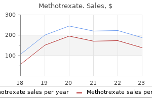
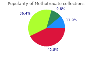
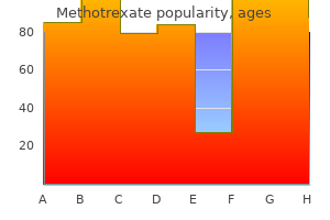
Methotrexate dosages: 10 mg, 5 mg, 2.5 mg
Methotrexate packs: 10 pills, 20 pills, 30 pills, 60 pills, 90 pills, 120 pills, 180 pills, 270 pills, 360 pills

A dorsal method through the primary net area, perpendicular to the online itself and between the first and second metacarpals, is to be most popular on anatomical grounds however may not all the time obtain enough drainage on its own. The mid-palmar area is greatest approached via a mix of a transverse distal palmar incision with an indirect longitudinal extension (McDonald et al 2011). They are likely to be confined by the hypothenar fascia and result in swelling of the ulnar side of the hand with some flexion and adduction of the little finger. They may be drained by an incision alongside the ulnar border of the space without significant threat of harm to underlying buildings. There are 10 identifiable compartments in the hand: four comprise the dorsal interossei, three contain the palmar interossei, and three contain the thenar and hypothenar muscular tissues and adductor pollicis. Any or all of these compartments could additionally be injured by a rise in intracompartmental stress. The absolute stress at which harm is brought on has not been decided however is assumed to be lower than that required in the lower limb. Injury could additionally be caused by blast, burn or crush, and by injection, whether or not of therapeutic substances at low strain or of business brokers at excessive pressure. Symptoms may be as non-specific as aching of the hand at rest or on exercise, or the hand could turn into painful with rising swelling and lack of motion within the digits. On examination, an intrinsic minus-type hand (claw hand) may be seen, with extension of the metacarpophalangeal joints and flexion of the proximal interphalangeal joints. Any suspicion of a compartment syndrome must be followed by prompt decompression. It is usually potential to decompress thoroughly via two dorsal longitudinal incisions over the second and fourth metacarpals, in addition to launch of the flexor retinaculum. The dorsal compartments are addressed instantly and the volar compartments opened deep by longitudinal incision. The palmar floor varieties a deeply concave carpal groove, accentuated by the palmar projection of the radial and ulnar borders.
The xiphisternal joint and xiphoid process are palpable at the inferior end of the sternum; the joint often lies at the stage of the ninth thoracic vertebra. Posteriorly, the free ends of the eleventh and twelfth ribs may be Autonomicplexusesinthethorax Cardiac plexus the cardiac plexus has a superficial component inferior to the aortic arch, lying between it and the pulmonary trunk, and a deep half between the aortic arch and tracheal bifurcation (Ch. The superficial half is formed by the cardiac branch of the left superior cervical sympathetic ganglion and the decrease of the two cervical cardiac branches of the left vagus. The deep half is shaped by the cardiac branches of the cervical and higher thoracic sympathetic ganglia and of the vagus and recurrent laryngeal nerves. Branches from the cardiac plexuses also kind the left and proper coronary and atrial plexuses. Pulmonaryplexus the pulmonary plexuses are anterior and poste rior to the other constructions at the hila of the lungs (Ch. They are shaped by cardiac branches from the second to fifth (or sixth) thoracic sympathetic ganglia and from the vagus and cervical sympathetic cardiac nerves. Vagal fibres either cross by way of the plexus or are given off instantly by the vagus within the thorax. All fibres relay within the oesophageal wall and are motor to the smooth muscle within the lower oesophagus and secretomotor to mucous glands in the oesophageal mucosa. Vasomotor sympathetic fibres arise from the higher six thoracic spinal twine segments. Those from the upper segments synapse in cervical ganglia; postganglionic axons innervate the vessels of the cervical and upper thoracic oesophagus. Fibres from the decrease segments pass on to the oesophageal plexus or to the coeliac ganglion, where they synapse; postganglionic axons innervate the vessels of the distal oesophagus. The accuracy of some older descrip tions of clinically necessary floor landmarks, based mostly on cadaveric or radiographic research, has been questioned lately (Hale et al 2010). The tail of the breast extends in the direction of the axilla along the infero lateral border of pectoralis major (Ch.
Diseases
The most common anastomoses are with the circumflex department of the left coronary artery by way of the posterior pericardial reflections and reflect the shut proximity of the bronchial arteries throughout the pulmonary hila. Extracardiac communications also exist with coronary atrial branches, especially the sinu-atrial nodal artery. Coronary angiography could additionally be performed by introducing a catheter through the femoral, radial or brachial arteries. The femoral artery is punctured with a needle three cm beneath the inguinal ligament while the leg is held adducted and barely externally rotated. The actual position is guided by palpation of the femoral arterial pulse, and the needle is inserted at an angle of 45�. After arterial puncture, a fantastic guidewire is inserted via the needle and fed into the artery. The catheter is then inserted over the guidewire and manipulated by way of the iliac artery into the aorta, up the aortic arch and into the ascending aorta. The brachial or radial artery could also be used for percutaneous entry to the circulation. Once the catheter is situated within the ascending aorta, quite so much of guidewires may be used to enter the coronary vessels for selective arteriography and interventions. Angiography is carried out with commonplace high-osmolality contrast medium with cineangiography. In chosen patients, new-generation, low-osmolality contrast medium may be used. The ostium of the left coronary artery arises from the left aortic sinus and is finest considered in the direct frontal and left anterior indirect projections. The right anterior oblique view is useful in demonstrating the diagonal branches and anterior interventricular (descending) coronary artery. The proper coronary artery originates from the best sinus of Valsalva and is often visualized in the proper anterior indirect view.
The connective tissues of these muscles and their attachment sites on the pectoral girdle innervated by the accessory nerve are derived from neural crest (Matsuoka et al 2005). Early limb development is patterned by somatopleuric mesenchyme, inside a permissive interplay with the limb ectoderm. Upper limb myoblasts migrate laterally from the ventrolateral border of cervical dermomyotomes and type dorsal and ventral lots prior to migration into the limb bud. After a short period within the limb bud, a subset of the myoblasts migrate medially out of the limb bud and into the axial mesenchyme to form the pectoral muscular tissues ventrally and latissimus dorsi dorsally. Only myoblasts undergo this second, medial, migration out of the limb bud; the somatopleuric mesenchyme stays inside the proximal limb anlagen and produces the glenohumeral joint and coracoid course of. Some myoblasts, originating from the ventrolateral edges of cervical dermomyotomes and extending because the hypaxial domain of the somite, swap off their myogenic destiny and observe a cartilage lineage to kind the medial a part of the scapula. The remaining hypaxial area myoblasts type serratus anterior, and the rhomboid muscles, deep pectoral girdle muscular tissues, attach to this medial part (Valasek et al 2011). The proximal and distal attachments of trapezius, to the nuchal line of the occipital bone and the higher a half of the backbone of the scapula, respectively, are derived from post-otic neural crest cells (Matsuoka et al 2005). The attachment of sternocleidomastoid to the clavicle also demonstrates its dual origin from neural crest cells and somatopleuric mesenchyme. The proximal portion, still displaying the dorsal bulge and ventral curve, is the shoulder and upper arm region; the next distal portion can now be recognized as the forearm; the most distal portion is now expanded into a flattened hand plate. Crenation of the hand plate between the digit rays continues in stage 18 embryos (44 days). Changes throughout phases 19�23 are involved with development of the limb and separation of the digits. The distal phalangeal parts of the fingers enlarge at stage 21, forming the nail beds.
This is normally considered to be an oncological emergency and symptoms are often utterly and promptly relieved by insertion of a vascular stent through the widespread femoral vein or by radiotherapy to the affected area after a tissue analysis is established. The thoracic part may be very brief, and is partly inside and partly exterior the pericardial sac. The extrapericardial part is separated from the proper pleura and lung by the proper phrenic nerve, and the intrapericardial half is roofed, except posteriorly, by inflected serous pericardium. Collateral venous channels In obstruction of the upper inferior vena cava, the azygos and hemiazygos veins and vertebral venous plexuses are the principle collateral channels that maintain venous circulation. They join the superior and inferior venae cavae and communicate with the widespread iliac vein by the ascending lumbar veins and with many tributaries of the inferior vena cava. Inferior vena cava the inferior vena cava returns blood to the center from infradiaphragmatic tissues. A left superior vena cava might have a slender connection with the right and then cross the left aspect of the aortic arch to cross anterior to the left pulmonary hilum earlier than turning to enter the proper atrium. It replaces the indirect vein of the left atrium and coronary sinus, and receives all of the tributaries of the coronary sinus. The left brachiocephalic vein typically tasks above the manubrium (more regularly in childhood), and crosses the suprasternal fossa in entrance of the trachea. A left-sided superior vena cava may cause difficulties when inserting a cardiac catheter, pacing or defibrillating electrodes as a end result of the angle between the left superior vena cava and the left subclavian vein is much more acute than that between the subclavian and a traditional left brachiocephalic vein. Even if insertion of a catheter into a left superior vena cava is possible, the angle at which the catheter enters the right atrium causes difficulty when making an attempt to place it into the proper ventricle and pulmonary trunk; this generally leaves the catheter tip in opposition to the coronary sinus, making it troublesome to obtain blood samples. In nearly all of persistent left superior venae cavae, blood drains into the best atrium by way of the coronary sinus. Cyanosis displays a persistent right-to-left shunt and affected people have the next danger for paradoxical embolism. If the superior vena cava is duplicated, the right superior vena cava might drain into the best atrium and the left superior vena cava into the left atrium. Embryologically, if the proper subcardinal vein fails to anastomose with the hepatic sinusoids, the hepatic phase of the inferior vena cava fails to develop.
Syndromes

The leaflet has a deep crescentic tough zone that receives numerous chordae tendineae. The ridge limiting the outer margin of the rough zone indicates the maximal extent of surface contact with the mural leaflet in full closure. Notice the thick wall of the left ventricle and the chordae tendineae of the mitral valve attaching to the papillary muscular tissues. Also shown are the continuity of the subaortic curtain with the mitral aortic leaflet. During passive ventricular filling and atrial systole, its easy atrial surface is important in directing a clean circulate of blood in path of the body and apex of the ventricle. After the onset of ventricular systole and closure of the mitral valve, the ventricular facet of its clear zone merges into the smooth surface of the subaortic curtain, which, with the remaining fibrous partitions of the subvalvular aortic vestibule, varieties the sleek boundaries of the ventricular outlet. Lack of definition of the major intervalvular commissures has led to disagreement and confusion regarding the territorial extent of this leaflet and the potential existence of accent scallops. Examination of the valve in the closed position reveals that the posterior leaflet might conveniently be regarded as comprising all the valvular tissue posterior to the anterolateral (inferoseptal) and posteromedial (superoposterior) ends of the most important zone of apposition with the aortic leaflet. Thus defined, it has a wider attachment to the anulus than does the anterior leaflet, guarding twothirds of the circumferential attachments. Further indentations normally divide the mural leaflet into a relatively large middle scallop and smaller lateral and septal commissural scallops. Each scallop has a crescentic opaque rough zone, receiving on its ventricular aspect the attachments of the chordae that outline the area of valvular apposition in full closure. Much extra of the mural leaflet is in apposition with the aortic leaflet during closure of the mitral valve. Precise papillary contraction, and rising pressure within the chordae, continue to prevent valvular eversion and preserve valvular competence. The orifices and the leaflets of each atrioventricular valves bear considerable modifications in position, form and area throughout a cardiac cycle.
Left coronary distribution is reciprocal and consists of most of the left ventricle; a slender strip of proper ventricle; the anterior two-thirds of the interventricular septum; and many of the left atrium. The term is deceptive as a outcome of the left artery virtually at all times supplies a greater quantity of tissue than the right. In more than 50% of individuals, the right atrium is provided solely by the right coronary artery, and in the the rest the availability is twin. Posterior ventricular branches are smaller and fewer as a result of the left ventricle is partly equipped by the posterior (inferior) interventricular artery. The artery to the sinu-atrial node is usually derived from the anterior circumflex section (less typically from the circum-marginal segment). The artery to the atrioventricular node, sometimes the terminal department of the circumflex artery (20%), arises near the crux. The broad variation in frequency signifies that many bridges could additionally be asymptomatic throughout life. The major scientific circumstances produced by a myocardial bridge are cardiac ischaemia, atherosclerosis and sudden cardiac demise. The incidence of atherosclerosis is increased when the proper coronary artery is bridged. Although a relationship between myocardial bridges and sudden cardiac dying has not been established, post-mortem series have shown histological proof of otherwise unexplained ischaemia in individuals with myocardial bridges; many died throughout train and had no different risk elements for coronary arterial illness. Arterial supply to the sinuatrial and atrioventricular nodes additionally varies: the sinu-atrial node is provided extra typically by the best coronary artery; fewer than 10% of sinu-atrial nodes obtain a bilateral supply. Acquired coronary artery fistulae are most commonly iatrogenic in aetiology but can also happen after traumatic harm; these mostly are of the coronary cameral type, from the proper coronary artery into the best facet of the center. Coronary anastomoses the cardiac collateral circulation represents a native system for coronary arterial bypass. The first few centimetres of the arterial main stems are devoid of anastomotic branches, however further distally, collateral channels are ample, exhibit variable calibres and occupy numerous areas, allowing for bidirectional flow between most native arteries. Anastomoses between branches of the coronary arteries, each subepicardial and myocardial, and between these arteries and extracardiac vessels, are of prime medical significance.
The superficial head arises from the distal border of the flexor retinaculum and the distal a half of the tubercle of the trapezium, and passes along the radial aspect of the tendon of flexor pollicis longus. It is connected by a tendon that accommodates a sesamoid bone to the radial aspect of the bottom of the proximal phalanx of the thumb. The deep part arises from the trapezoid and capitate bones and from the palmar ligaments of the distal row of carpal bone, and passes deep to the tendon of flexor pollicis longus. It Flexor pollicis brevis Testing the affected person abducts the thumb at proper angles to the palm in opposition to resistance; the muscle can be seen and felt. It arises from the tubercle of the trapezium and the flexor retinaculum, and is attached to the whole length of the lateral border, and the adjoining lateral half of the palmar floor of the metacarpal bone of the thumb. The deep layer is shown after slicing the flexor retinaculum and partial removing of several superficial muscular tissues: palmar aspect. The oblique head is attached to the capitate bone, the bases of the second and third metacarpal bones, the palmar ligaments of the carpus, and the sheath of the tendon of flexor carpi radialis. Most of the fibres converge right into a tendon (containing a sesamoid bone) that unites with the tendon of the transverse head and is connected to the ulnar side of the bottom of the proximal phalanx of the thumb. The deepest fibres might pass into the medial side of the dorsal digital growth of the thumb. The fibres converge to be attached, with the oblique head and the primary palmar interosseous, to the bottom of the proximal phalanx of the thumb. Adductor pollicis Relations the deep palmar arch and the deep branch of the ulnar nerve pass between the two heads of the muscle. Anteriorly, adductor pollicis is crossed by the flexor tendons of the index finger and their sheath, and the primary lumbrical, and is overlapped by flexor pollicis brevis. Posteriorly, it abuts against the first dorsal interosseous muscle; together, these muscular tissues form the mass of the first web area of the hand. Actions Adductor pollicis is the biggest and strongest of the intrinsic muscles and acts to approximate the thumb to the palm of the hand.
The epithelium within the respiratory bronchioles progressively reduces in peak in direction of the alveoli, and is finally composed of cuboidal, non-ciliated cells. Respiratory bronchioles have lateral pouches in their walls, which are lined with squamous cells, so offering an accessory respiratory floor. Lymphocytes and mast cells migrate into the epithelium from the underlying connective tissue. Clusters of lymphocytes generally lie beneath nonciliated epithelial cells of the microfold (M-cell) sort. They resemble connective tissue mast cells, and their cytoplasmic histaminecontaining granules are launched in response to irritants, together with inhaled allergens. Submucosal glands Tubuloacinar, seromucous glands are current within the submucosa of the trachea and bronchi and, to a lesser extent, within the larger bronchioles. They comprise separate mucous and serous cells and are an essential source of the mucus on the surface of the ciliated respiratory epithelium. Their secretions embody mucins; bacteriostatic substances similar to lysozyme and lactoferrin; secretory antibodies (immunoglobulin A (IgA)) produced by plasma cells within the submucosal connective tissue; and protease inhibitors. Deficiency of 1-antitrypsin causes continual obstructive pulmonary illness, by inducing panacinar emphysema and bronchiectasis. The secretory acini and tubules are surrounded by myoepithelial cells, which are innervated by autonomic fibres (see above). Ciliated columnar cells Goblet cells Goblet cells are current from the trachea (6000�7000 per mm2) distal to the smaller bronchi, but are normally absent from bronchioles. They comprise an apical area full of huge secretory vacuoles filled with mucinogen. Clara cells Clara cells are cuboidal, non-ciliated cells with apices that bulge into the lumen. Basal cells contact the basal lamina and are most frequent in the larger conducting passages. The cilia extend right into a watery fluid secreted by serous cells of the submucosal glands, but their tips are involved with a extra superficial layer of thicker mucus secreted by surface goblet cells and mucous cells in the submucosal glands. The price of ciliary beating is often 12�16 per second; mechanical stimulation of the epithelial surface and inflammatory mediators increase the speed. In addition to tight junctions, which seal the apical intercellular space from the airway lumen, the ciliated cells are coupled by gap junctions, which permit a change in fee of beating to spread from stimulated cells to their neighbours (probably through calcium signalling) so that their metachronal coordination remains intact.
Its fusiform muscular belly converges in the mid-forearm into an extended tendon that passes inside a synovial sheath through a lateral canal, shaped by the flexor retinaculum above and a groove on the trapezium beneath. It inserts on the palmar surface of the bottom of the second metacarpal and sends a slip to the third metacarpal. Distally, it may also be attached to the flexor retinaculum, trapezium or fourth metacarpal. It acts with flexor carpi ulnaris to effect balanced wrist flexion, and in concert with the radial extensors of the wrist to achieve balanced abduction of the hand. Testing Flexor carpi radialis is tested by palpating its contracting fibres during flexion of the wrist in opposition to resistance. The humero-ulnar head arises from the medial epicondyle of the humerus via the frequent flexor origin, the anterior band of the ulnar collateral ligament, adjacent intermuscular septa, and from the medial facet of the coronoid process proximal to the ulnar origin of pronator teres. The radial head, a skinny sheet of muscle, arises from the anterior radial border between the radial tuberosity and the insertion of pronator teres. The superficial stratum, joined laterally by the radial head, divides into two tendons for the middle and ring fingers. The deep stratum offers off a muscular slip to be a part of the superficial fibres directed to the ring finger, and ends in two tendons for the index and little fingers. The 4 tendons diverge Relations At its origin, flexor carpi radialis lies medial to pronator teres. In the decrease part of the forearm, the radial artery lies between the tendon of flexor carpi radialis and the tendon of brachioradialis; the radial pulse may be palpated proximal to the wrist between these two tendons. Vascular provide Flexor carpi radialis is equipped by a single dominant proximal pedicle and several other distal minor pedicles. The dominant pedicle is shaped by a branch that arises from both the anterior or the posterior ulnar recurrent artery. The posterior ulnar recurrent artery passes deep to pronator teres to enter the deep floor of flexor carpi radialis; from right here, it divides right into a small ascending department and a bigger descending department.
Basir, 46 years: Oesophageal glands Groups of small tubule-acinar oesophageal glands mainly lie in the submucosa, each sending a single lengthy duct through the intervening layers into the lumen. The former is distal on the anterior scaphoid floor and palpable (sometimes, also visible) as a small medial knob at the proximal border of the palmar thenar eminence, radial to the tendon of flexor carpi radialis.
Luca, 28 years: The true chordae often come up from small projections on the tips or margins of the apical third of papillary muscles, though they sometimes arise from the papillary muscle bases or directly from the ventricular partitions and septum. The inferior aorticopulmonary body is close to the center and anterior to the aorta, and the center aorticopulmonary physique is close to the proper aspect of the ascending aorta.
References
Realice búsquedas en nuestra base de datos:
