

Francisco Badosa MD, FACS
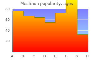
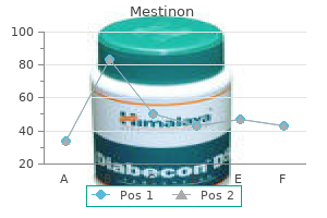
Mestinon dosages: 60 mg
Mestinon packs: 30 pills, 60 pills, 90 pills, 120 pills, 180 pills, 270 pills
Both scanning electron microscopy evaluation and transmission electron microscopy analysis allow the lower particle sizing limit to be greatly extended over that possible with a light microscope. Sample preparation and evaluation conditions Particle measurement distributions can be determined by examination of a powder because it sediments in a liquid. If the powder is hydrophobic, it might be necessary to add a dispersing agent to aid wetting. In instances where the powder is soluble in water, it is going to be essential to use nonaqueous liquids or perform the evaluation in a gasoline. Image evaluation With manual microscopy, just a few particles could be examined and sized in an affordable time. This dangers number of an unrepresentative pattern, operator subjectivity and operator fatigue. Automated image evaluation, for mild and electron microscopy, has the advantages of being extra goal and much sooner than guide analysis, and it additionally enables a a lot wider variety of size and form parameters to be processed. Image analysis could also be static, whereby particles on a microscope slide are inspected with use of a microscope and digital camera, or dynamic (flowimage analysis), whereby pictures of particles dispersed in a liquid are captured by a digital camera as they move by way of a circulate cell. Principles of measurement the methods of dimension analysis by sedimentation can be divided into two major classes based on the tactic of measurement used. One kind is based on the measurement of particles in a retention zone; a second sort makes use of a nonretention measurement zone. An instance of a nonretention zone measurement technique is named the pipette methodology. In this methodology, identified volumes of suspension are withdrawn and the focus variations are measured with respect to time. The Andreasen fixed-position pipette comprises a graduated cylinder which may hold approximately 500 mL of suspension fluid. A pipette is positioned centrally within the cylinder and is held in place by a ground-glass stopper so that its tip coincides with the zero stage.
Gluteus maximus and tensor fasciae latae gain attachment to the tract and, due to this attachment, assist in extension and stabilization of the knee. It is attached to bone across the margins of the patella, to the medial surface of the tibia and, inferiorly, to each malleoli. It ensheaths the muscular tissues and contributes to intermuscular septa that separate the anterior, lateral and posterior muscle compartments. The posterior muscles are further divided by a fascial envelope into superficial and deep compartments, the former enclosing gastrocnemius and soleus. Around the ankle the fascia thickens to kind the retinacula, restraining the tendons as they enter the foot. Throughout the leg and thigh, the fascia is pierced by perforating (communicating) veins becoming a member of the superficial to the deep veins, and by cutaneous nerves, arteries and lymph vessels. The fascial compartments of the leg are quite inflexible envelopes for the gentle tissues contained within them; trauma and bleeding, such as may comply with a leg fracture, may cause compartmental swelling and rise in stress of adequate severity to compromise the arterial supply to the contents of the compartment. It is essential in these circumstances that close remark is maintained on the pulses within the leg, and if the circulation is found to be threatened, a fasciotomy (incision along the length of the fascial sheath) must be urgently undertaken to relieve the pressure in that compartment. Valves are current in the bigger veins and all of the communicating veins; they direct blood circulate in the course of the heart or from the superficial to the deep veins. The superficial veins drain skin and superficial fascia into two major channels, the good and small saphenous veins. It receives tributaries from the small saphenous vein and connects by speaking branches to the deep veins of the thigh and calf simply behind the medial border of the tibia. It receives cutaneous tributaries and communicates with the deep veins of the calf by perforating veins. The deep veins comprise those of the foot and the soleal plexuses of veins, the popliteal and the femoral vein (p. The superficial veins drain to them by speaking veins that perforate the deep fascia. All the veins of the lower limb possess valves that let blood circulate solely up the limb or from superficial veins to deep veins.
Diseases
Even the original posterior border of stomach grows faster, forming the higher curvature. The stomach also rotates along anteroposterior axis, in order that distal or pyloric half moves to right and proximal or cardiac part moves to left side. The 90� rotation of abdomen along the vertical axis pulls the dorsal mesogastrium to the left side creating the lesser sac or omental bursa. Spleen seems as mesodermal condensation in the left leaf of dorsal mesogastrium. The sympathetic nerves to the stomach are equipped by segments T6�T9 of the spinal wire, which additionally provide the upper part of the belly walls so the ache of gastric ulcer is referred to epigastric region. The ache of foregut-derived areas is referred to epigastric region; those of midgut-derived organs to periumbilical area whereas those of hindgut-derived viscera to the suprapubic area. A review that discusses the precept anatomical mechanisms that prevent reflux on the gastro-oesophageal junction and explains how failure of a quantity of of those might result in progressive gastrooesophageal reflux illness. A posteriorly perforating peptic ulcer will most probably produce peritonitis within the following: a. Intestines suffer from bacterial infection like typhoid, tuberculosis; parasitic infection, like roundworm, tapeworm, etc. The proximal 1� elements of duodenum, together with liver, gallbladder and pancreas, develop from foregut. The distal 2� components of duodenum, jejunum, ileum, caecum, appendix, ascending colon and proper two-thirds of transverse colon develop from midgut. Lastly, the left one-third of transverse colon, descending colon, pelvic colon and proximal part of rectum develop from hindgut. The length is larger in males than in females, and higher in cadavers, as a outcome of loss of tone than in the dwelling. It is split into: 1 An upper, fixed half, known as the duodenum, which measures about 25 cm in length; and 2 A lower, cell part, forming a really lengthy convoluted tube. The circular folds of mucous membrane, plicae circulares, or valves of Kerckring kind complete or incomplete circles.
It accompanies the subcostal artery alongside the lower border of 12th rib and passes behind the lateral arcuate ligament. It lies behind the kidney, anterior to quadratus lumborum, pierces the aponeurosis of 511 the intercostal and subcostal nerves and their collateral branches provide intercostal muscular tissues and muscles of anterolateral abdominal wall. Cutaneous the terminal components of T7�T12 nerves are referred to as the anterior cutaneous branches. T10 supplying the pores and skin round umbilicus; T7, the skin of epigastrium and T8, T9, the intervening pores and skin between epigastrium and the umbilicus. T11, T12 and iliohypogastric (L1) provide the pores and skin between umbilicus and pubic symphysis. The lateral cutaneous branches of the T7�T11 intercostal nerves divide into anterior and posterior branches to supply the pores and skin of lateral aspect of abdomen and back. The lateral cutaneous branch of T12 provides the pores and skin of upper anterior part of the gluteal area. The dorsal divisions of those rami give rise to lateral cutaneous nerve of thigh (L2, 3), and femoral nerve (L2, three, 4) (Appendix 1). Branches arising from ventral divisions are: 1 Nerve to quadratus femoris (L4, 5, S1): Supplies quadratus femoris, inferior gemellus and hip joint. Branches from dorsal divisions are: 1 Superior gluteal nerve (L4, 5, S1): Supplies gluteus medius, gluteus minimus and tensor fascia latae. Muscular branches to deep transversus perinei, compressor urethrae, sphincter urethrovaginalis, ischiocavernosus, bulbospongiosus, external anal sphincter, levator ani, corpus spongiosum, penis and urethra, lower 2. Only higher two ganglia receive white ramus communicans from the ventral primary rami of first and second lumbar nerves. Branches Abdomen and Pelvis Pudendal Nerve Pudendal nerve supplies the pores and skin, external genital organs and muscle tissue of perineum. It is worried with micturition, defaecation, erection, ejaculation and in females, with parturition. These move alongside the spinal nerves to be distributed to the sweat glands, cutaneous blood vessels and arrector pili muscle tissue (sudomotor, vasomotor and pilomotor). Course It begins within the pelvis, enters the gluteal region via greater sciatic notch, lies on the sacrospinous ligament, leaves the gluteal area via lesser sciatic notch.
The chain bears four sacral ganglia on all sides and the only ganglion impar within the central part. It covers the lateral pelvic wall and the pelvic flooring known as parietal pelvic fascia; and in addition surrounds the pelvic viscera referred to as visceral pelvic fascia. Principles of Distribution 1 the fascia is dense and membranous over nonexpansile structures. In this respect, the fascia of Waldeyer is an exception, which extends from the sacrum to the ampulla of rectum. It is hooked up alongside a line from iliopectineal line to the inferior border of pubic bone. Because of the loose nature of the fascia, infections can unfold rapidly inside it. The varied ligaments are dealt with individual viscera together with the prostate, bladder, uterus and the rectum. Visceral Pelvic Fascia this fascia surrounds the extraperitoneal elements of the pelvic viscera. It is unfastened and mobile around distensible organs like bladder, rectum and vagina, but is dense around non-distensible organs, just like the prostate. The visceral layer is connected alongside a line extending from the middle of back of pubis to the ischial spine. The levator ani and coccygeus may be thought to be one morphological entity, divisible from before backwards into the pubococcygeus, the iliococcygeus and the ischiococcygeus or coccygeus. They have a steady linear origin from the pelvic floor of the body of the pubis, the obturator fascia or white line or tendinous arch and the ischial spine. The muscle fibres slope downwards and backwards to the midline, making a gutter-shaped pelvic floor. Pubococcygeus Part these fibres surround the vagina and kind the sphincter urethrovaginalis. Iliococcygeus Part Abdomen and Pelvis 1 the anterior fibres of this half come up from the medial a part of the pelvic surface of the physique of the pubis. In the male, these fibres intently encompass the prostate and constitute the levator prostatae.
Larger spaces (cisterns) are present the place the arachnoid stretches across concavities of the brain; these embrace the cerebellomedullary cistern (cisterna magna) within the angle between the cerebellum and medulla, the interpeduncular cistern between the cerebral peduncles and the pontine cistern in front of the pons. The lumbar cistern, caudal to the end of the spinal cord, contains the cauda equina. The subarachnoid house extends alongside the bundles of the olfactory nerve that penetrate the cribriform plate, and alongside the optic nerve as far as the optic disc within the eyeball. Blows to the head cause sudden extreme deceleration, producing extreme drive on the cranial contents. There are ill-understood adjustments inside the brain that trigger a sudden abrupt loss of consciousness instantly after the blow to the top (cerebral concussion). In the absence of any seen structural injury and intracranial haemorrhage this loss of consciousness is short-lived. However, intracranial haemorrhage is associated with a significant variety of often extra extreme head accidents. Extradural haemorrhage between the two layers of dura is usually the outcomes of bleeding from the center meningeal artery, which is incessantly associated with fractures of the temporal bone. This is arterial bleeding and the blood accumulates fairly rapidly, inflicting compression of the mind and a gradual loss of consciousness. Surgical evacuation of the haematoma and stopping of the haemorrhage is normally achieved via a burr hole drilled within the pterion (p. It is commonest in elderly people, in whom some shrinkage of the mind has occurred. The damage could additionally be apparently quite trivial but the smaller mind allows it to be shaken within the skull, which is followed by a tear in a cerebral vein as it enters the superior sagittal sinus. The venous bleeding is slower than that of arterial trauma, and the outcomes of accelerating cerebral compression are thus slower in onset. It leads to in depth meningeal irritation as the blood spreads beneath the meninges to cause headache and neck stiffness, progressing to eventual lack of consciousness. Its complete volume is about one hundred thirty mL, of which one hundred mL is in the subarachnoid house and 30 mL within the ventricles.
Vidhara (Hawaiian Baby Woodrose). Mestinon.
Source: http://www.rxlist.com/script/main/art.asp?articlekey=96344
The squamous half, a flat plate, extends backwards and upwards to articulate with the parietal and mastoid part of the temporal bone. Lymph vessels cross to lymph nodes in the round chain around the base of the skull; posteriorly to the occipital and mastoid nodes and anteriorly to the preauricular nodes. In males, facial hair is very dense over the temporal fossa, the zygomatic arch and the mandibular region. The mucocutaneous junction around the mouth is on the facial facet of the lips (the red margin). Mucous and small salivary glands are current on the inner facet of the lips and cheeks. Orbicularis oris is the sphincter around the mouth and varieties the greater a part of the substance of the lips. Other muscular tissues blend with it, such as the buccinator and the levator and depressor muscle tissue, that are connected to the angles of the mouth (anguli) and to the middle of the lips (labii). It has a continuous lateral attachment to the pterygomandibular raph� and the outer surfaces of the maxillae and mandible adjacent to the final molar tooth. Its fibres cross forwards and medially, Frontalis Corrugator Orbicularis oculi Levator labii superioris Levator labii superioris alaeque nasi Levator anguli oris Zygomaticus main and minor Masseter Buccinator Orbicularis oris Depressor anguli oris Mentalis Depressor labii inferioris Platysma (partly removed superiorly) Thick Skin containing many hair follicles and sweat glands. Musculofibrous epicranial Aponeurosis containing the occipitalis muscle posteriorly and the frontalis muscle in front. Anteriorly the frontalis has no bony attachment but blends with the fibres of the orbicularis oculi muscle tissue. Loose areolar tissue, which allows free movement of the aponeurosis and overlying skin over the periosteum. Scalping accidents, such as these brought on by lengthy hair catching in shifting equipment, pull the outer layers of the scalp away from the cranium alongside this layer. The Periosteum (pericranium) of the bones of the vault, which is steady by way of the sutures of the vault with the endocranium.
The central branches perforate the white matter to supply the thalamus, the corpus striatum, and the inner capsule. Central Branches Anteriorly: Anterior communicating artery becoming a member of the 2 anterior cerebral arteries. Posterolaterally: Posterior communicating arteries Posteriorly: Posterior cerebral arteries the circulus arteriosus makes an attempt to equalize the flow of blood to totally different elements of mind and supplies a collateral circulation in the event of obstruction to one of its elements. There is hardly any mixing of bloodstreams on proper and left sides of the circulus arteriosus. These enter the medialmost part of anterior perforated substance to provide preoptic and supraoptic regions of anterior hypothalamus. These divide in two sets: Medial striate ascends by way of lentiform nucleus to provide this nucleus including caudate nucleus and internal capsule. The lateral striate ascends lateral to lentiform nucleus, flip medially, pass via the substance of this nucleus to enter inside capsule. These supply tuberoinfundibular and mammillary areas of hypothalamus, subthalamus, anterior and medial elements of thalamus, medial a part of tegmentum and crus cerebri of midbrain. Inferior temporal gyrus excluding the a half of the temporal pole can be provided by posterior cerebral artery. Two arteries are related by the anterior speaking artery Winds round cerebral peduncle to reach the tentorial surface of cerebrum 1. Cerebral Cortex Cerebral cortex is equipped by branches of all three cerebral arteries. Intimately applied to the capillaries, there are numerous processes of astrocytes and it has been estimated that these processes cover about 80% of the capillary surface. These embrace pineal physique, hypophysis cerebri, choroid plexuses and area postrema in fourth ventricle of brain. The chief inner supply of autoregulation is the adjustment of arterial muscle tone in response to intraluminal stress adjustments. Cerebral blood move remains at 60�70 ml/100 g/min during systemic blood stress; adjustments ranging from 80 to 180 mm Hg. This is achieved by a direct myogenic response to distension produced by rising intraluminal stress. The H+ ion focus within the perivascular area is the chief external supply of autoregulation of cerebral blood vessels.
Clean the superior mesenteric vessels with its branches both from its right and left surfaces. These happen on the aspect of the mesenteric border at the website of entry of vasa recta. Vulnerability of jejunum is as a result of of relative weak point of the longitudinal muscle coat. It is usually adopted by a rapidly spreading type of intestinal obstruction because of the haemorrhagic infarction of the concerned gut. This part of duodenum may get obstructed as it lies between abdominal aorta and superior mesenteric artery. Origin 2 Inferior mesenteric artery arises from the entrance of the stomach aorta behind the third a part of the duodenum, at the degree of third lumbar vertebra, and three to 4 cm above the bifurcation of the aorta. Opposite third sacral vertebra, it divides into proper and left branches which descend one on all sides of the rectum. They pierce the muscular coat of the rectum and divide into a quantity of branches, which anastomose with each other at the level of the anal sphincter to form loops around the lower finish of the rectum. Left Colic Artery 1 the inferior mesenteric vein drains blood from the rectum, the anal canal, the sigmoid colon and the descending colon. The superior rectal vein crosses the left frequent iliac vessels medial to the left ureter and continues upwards as the inferior mesenteric vein. The vein ascends behind the peritoneum, passes lateral to the duodenojejunal flexure in the free margin of paraduodenal fold and behind the physique of the pancreas. They pass downwards and to the left, and anastomose with one another to form the decrease a part of the marginal artery. The uppermost department anastomoses with the descending department of the left colic artery, whereas the bottom sigmoid artery sends a department to anastomose with the superior rectal artery. Marginal artery was described by von Haller in 1803 and its current name was given by Sudeck in 1907. The marginal artery is an arterial arcade located alongside the concavity of the colon. It is shaped by anastomoses between the main arteries supplying the colon, namely the ileocolic, proper colic, center colic, left colic and sigmoid arteries. The marginal artery is able to supplying the colon even within the absence of one of the main feeding trunks.
Pelvic splanchnic nerves give fibres to inferior hypogastric plexuses to supply rectum and higher half of anal canal. Some of inferior hypogastric plexuses move up through superior hypogastric plexus and get distributed along the branches of inferior mesenteric artery to the left third of transverse colon, descending and sigmoid colons. Rectum and Anal Canal Sympathetic provide reaches from coeliac plexus alongside gastric and gastroepiploic arteries. The left vagus types anterior gastric, whereas proper vagus contains posterior gastric nerve. The anterior gastric nerve provides cardiac orifice, anterior floor of body as nicely as fundus of stomach and pylorus. Posterior gastric nerve supplies posterior floor of body and fundus till pyloric antrum. Small Intestine Sympathetic fibres move alongside inferior mesenteric and superior rectal arteries and also through superior and inferior hypogastric plexuses. Parasympathetic supply is from pelvic splanchnic nerves, which join inferior hypogastric plexus. Afferent impulses of physiological distension of rectum and sigmoid colon are carried by parasympathetic nerves, whereas ache impulses are conveyed both by the sympathetic and parasympathetic nerves. Pancreas Brain�Neuroanatomy the nerves of this a part of the gut are derived from coeliac ganglia fashioned by posterior gastric nerve (vagus) and splanchnic nerves (sympathetic) and the plexus around superior mesenteric artery. Parasympathetic system is secretomotor to the intestines and inhibits the sphincters. The nerve fibres make synaptic contact with acinar cells before innervating the islets. The parasympathetic ganglia lie in sparse connective tissue of the gland and the islet cells. Liver Nerves of the liver are derived from hepatic plexus which is an offshoot of coeliac plexus.
Gamal, 29 years: Third Neuron Third neuron lies in the posterolateral ventral nucleus of the thalamus. Superior and inferior ophthalmic veins drain the orbit and cross via the superior orbital fissure to the cavernous sinus. Its branches form components of the testicular, inferior mesenteric, iliac and superior hypogastric plexuses, and supply the inferior vena cava.
Rocko, 39 years: It is split into: 1 An higher, fixed half, known as the duodenum, which measures about 25 cm in size; and a pair of A lower, mobile half, forming a really long convoluted tube. It regulates ranges of awareness and some emotional elements of sensory experiences. Quadriceps is a big muscle mass forming the majority of the anterior region of the thigh.
Kaffu, 23 years: In the upright position of the physique, the venous return from the decrease limb relies upon largely on the contraction of calf muscle tissue. It originates on the posterior wall of the third ventricle and passes by way of the midbrain between the dorsal tectum and ventral tegmentum to enter the superior aspect of the fourth ventricle. S2, S3 and S4, to the hypogastric plexus beneath the aortic bifurcation, and its branches.
Mezir, 26 years: Anterior Spinal Artery It is formed by the union of a branch from every vertebral artery on ventral surface of medulla oblongata close to the pons. There are about 5 perforators along the nice saphenous vein, and one perforator alongside the small saphenous vein. Reflect this ligament from its proximal attachment to see the deeper plantar calcaneocuboid ligament.
Tarok, 60 years: The septal area can affect behaviour via connections to the hypothalamus and the brainstem. Gliding � lateral gliding separates the cartilages, abducts the vocal folds and produces an inverted V-shaped rima glottidis. Materials such as carbon black (a very finely divided form of carbon) have extremely large floor areas, and as such are wonderful adsorbents, both from resolution.
Iomar, 28 years: Presence of valves which help the long column of blood and divide the long column into shorter parts. Identify the parietal peritoneum, adherent to the parieties or partitions of the belly cavity. Parasympathetic stimulation produces pupillary constriction; sympathetic stimulation produces pupillodilatation.
Jorn, 43 years: In the vertebral column: a the person vertebrae are all separately identifiable within the adult b cervical vertebrae all have bifid spines c all thoracic vertebrae have articular surfaces for articulation with ribs d the vertebral our bodies bear articular processes arising close to the base of their pedicles e within the grownup the first fetal curvatures are retained within the thoracic and sacral regions 2. The contralateral artery can be demonstrated because the materials is introduced underneath strain and a few passes retrogradely right down to the other aspect: 1, vertebral arteries in transverse foramina; 2, vertebral arteries on basiocciput; three, basilar artery; four, superior cerebellar artery; 5, posterior cerebral artery. The internal structure of the medulla could be considered as being the dorsal tegmentum and the ventral pyramids.
References
Realice búsquedas en nuestra base de datos:
